MRI has become a beneficial tool in revealing how different parts of the brain are connected to each other and are related to autism spectrum disorder (ASD), according to a meta-analysis in the Harvard Review of Psychiatry.
The review from University of Massachusetts Medical School identified 33 functional MRI studies of ASD and reflected on a wide range of patient populations and study methods.
Most of the studies found "some amount of either reduction or loss of local or long-distance connectivity" associated with ASD, David Kennedy, PhD, and colleagues wrote (Harv Rev Psychiatry, July/August 2015, Vol. 23:4, pp. 223-244).
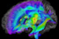
Thirty-six studies looked at structural connectivity of the brain in ASD. The findings were more consistent than the functional MRI studies, showing evidence of decreased connectivity in many areas of the brain's white matter.
About half of functional MRI studies tried to relate the functional connectivity findings to behavioral measures of ASD. Although many brain areas with reduced functional connectivity are known to be involved in the relevant behaviors, their true link to behavior in ASD is still unknown.
"The addition of behavioral correlates to the structural or functional connectivity studies would be immensely valuable in order to distinguish which of the many structural and functional changes that have been identified have specific behavioral consequences," according to Kennedy.
The next challenge is to generate more precise results and to determine whether those results can be correlated with the diverse profiles of patients with autism, the researchers concluded.


.fFmgij6Hin.png?auto=compress%2Cformat&fit=crop&h=100&q=70&w=100)


.fFmgij6Hin.png?auto=compress%2Cformat&fit=crop&h=167&q=70&w=250)


