Dear AuntMinnie Member,
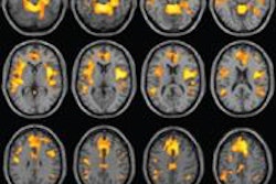
Have you ever wondered what sex looks like on MRI? Dutch researchers had that very thought, so they performed what's believed to be the first study of human coitus inside an MRI magnet.
That was way back in 1999, and the study's publication in BMJ that year raised eyebrows and even some criticism from those who thought the findings were unsuitable for publication. Still, the research offered a fascinating look at the anatomical processes involved in one of the most basic human urges.
The controversial study was revisited this month by lead researcher Dr. Pek van Andel, a physiologist at the University of Groningen, who reflected on the research and what we learned from it in a lecture in London organized by the British Institute of Radiology. Contributing writer Becky McCall was there to report for our sister site AuntMinnieEurope.com -- read all about it by clicking here.
CDS debate at AHRA
Is clinical decision-support (CDS) software a blessing or a curse? That was the question being discussed this week at the AHRA meeting in Las Vegas.
Liz Quam, executive director of the Center for Diagnostic Imaging's Quality Institute, pondered the pros and cons of CDS, which is designed to reduce inappropriate utilization of medical imaging exams. The hope is that the software will be less onerous than radiology benefits management (RBM) firms, which require preauthorization before some imaging exams can be performed.
Clinical decision-support software has the potential to be more flexible and less burdensome than RBMs, and a pilot study reported that it could also save money. But there's still much work to be done before CDS is adopted widely. Learn more by clicking here, or visit our Imaging Leaders Community at leaders.auntminnie.com.
Printing 3D hearts
Finally, visit our Advanced Visualization Community for a fascinating article on work by researchers from Massachusetts General Hospital, who are using 3D printing technology to create models of cardiac anatomy from CT scans.
The models can be used for a number of applications, from surgical planning to regenerative medicine. Find out how they do it by clicking here, or visit the community at av.auntminnie.com.


