If there was ever any doubt about the existence of the Christmas spirit, one need only look into certain regions of the human brain. That's according to Danish researchers, who found that specific areas of the brain lit up on functional MRI (fMRI) scans when people who celebrate the holiday viewed Christmas-related imagery.
The study, published online December 17 in BMJ, showed five areas of heightened brain activity in people who celebrate Christmas and its traditions, compared with those who have neutral feelings toward the holiday season.
"Understanding how the Christmas spirit works as a neurological network could provide insight into an interesting area of human neuropsychology and be a powerful tool in treating ailments such as bah humbug syndrome," wrote the group led by Anders Hougaard, a research fellow in neurology from Rigshospitalet and the University of Copenhagen (BMJ, December 17, 2015).
Christmas present
Functional MRI, of course, has been used for more than two decades in neuropsychological studies to explore emotional and functional centers of the brain and illustrate the sources of feelings such as joy and sorrow. But, arguably, never has the modality been utilized for such lofty, heavenly pursuits.
For the study, the researchers recruited 20 healthy control subjects who had been participating in another fMRI clinical trial. All subjects had normal or corrected-to-normal vision and had consumed no eggnog or gingerbread prior to their scans.
Half of the volunteers were placed in a "Christmas group"; these individuals celebrated the holiday according to Danish tradition. The other 10 subjects did not celebrate Christmas and formed the "non-Christmas group," which included Pakistani, Indian, Iraqi, or Turkish expatriates or people of Pakistani descent who were born in Denmark.
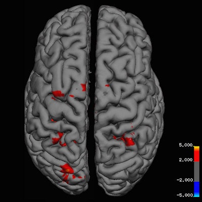
 Danish researchers identified areas of heightened brain activity in people who celebrate Christmas, compared with those who have neutral feelings. All images courtesy of BMJ.
MR images were obtained on a 3-tesla scanner (Achieva, Philips Healthcare), with T1-weighted magnetization-prepared rapid gradient-echo (MPRAGE) imaging for anatomical reference. Functional MRI scans used an echo-planar imaging sequence, along with cerebral perfusion through an arterial spin-labeling (ASL) sequence.
The fMRI protocol also measured blood oxygen level-dependent (BOLD) response while participants viewed a series of images. Some images had Christmas themes, while others were holiday-neutral.
The images were shown for two seconds at a time. After six consecutive images with a Christmas theme appeared, six everyday images were displayed.
Following their scans, all participants were asked to fill out a questionnaire about their ethnicity, Christmas traditions, and feelings associated with the holiday.
True believers
Baseline perfusion scans recorded normal results for all subjects and no significant differences between the two groups (p = 0.26). Both groups also reacted with increased brain activity in the primary visual cortex when shown Christmas images versus everyday scenes (p < 0.001).
However, fMRI detected significant increases in neural activations in the primary somatosensory cortex of subjects in the Christmas group when they viewed a holiday theme.
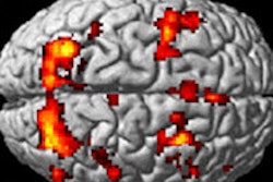
In fact, fMRI activation maps reflected five areas of heightened activity in response to holiday images in the Christmas group versus the non-Christmas group. These areas were the left primary motor and premotor cortex, right inferior and superior parietal lobule, and bilateral primary somatosensory cortex (p < 0.001).
"The left and right parietal lobules have been shown in earlier fMRI studies to play a determining role in self-transcendence, the personality trait regarding predisposition to spirituality," the authors wrote.
In addition, the frontal premotor cortex is important for experiencing emotions shared with others by mirroring or copying their body state, and premotor cortical mirror neurons even respond to observation of ingestive mouth actions.
There were no areas of the brain where the non-Christmas group had significantly greater reactions to Christmas images than the Christmas group.
Magically complex
Given the observed areas of cerebral response, the activity "coincided well with our hypothesis that images with a Christmas theme would stimulate centers associated with the Christmas spirit," the authors wrote.
Hougaard and colleagues noted some limitations of the research, such as a study design that did not determine if the increased brain activity was "Christmas-specific or the result of any combination of joyful, festive, or nostalgic emotions in general."
"Bringing these issues up, however, really dampened the festive mood," they added. "Therefore we, in the best interest of the readers of course, decided not to ruin the good Christmas cheer for everyone by letting this influence our interpretation of the study."
They did recommend further research -- presumably again with no eggnog or Christmas cookies prior to fMRI scans -- to confirm more definitively that the Christmas spirit is indeed in our heads. Research could also be extended to other holidays and commemorations, such as Easter, Hanukkah, or Diwali.
Another research possibility would be an imaging study to determine if the Grinch's heart was indeed two sizes too small, and what effect that might have on cardiac function.
"Although merry and intriguing, these findings should be interpreted with caution," the authors wrote.


