Many older patients recover more slowly from concussions than younger individuals, based on different brain activation patterns seen on functional MRI (fMRI) scans. The study results, published online October 6 in Radiology, suggest that mild traumatic brain injury (TBI) may have a more profound and lasting effect among older people.
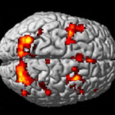
Initial fMRI scans one month after injury showed that younger patients with mild TBI demonstrated hyperactivation, or greater than normal brain activity, during memory tasks, while older patients had hypoactivation, or less than normal brain activity. Six-week follow-up scans revealed that the hyperactivation declined among the young patients, but hypoactivation remained among the older individuals.
"Taken together, these findings provide evidence for differential neural plasticity across different ages, with potential prognostic and therapeutic implications," wrote lead author Dr. David Yen-Ting Chen and colleagues from Shuang-Ho Hospital and Taipei Medical University in New Taipei City, Taiwan (Radiology, October 6, 2015).
Mild TBI happens
Memory problems have long been associated with the aftermath of concussions, which account for some 75% of all traumatic brain injuries. Neuropsychologic tests, CT, and MRI have not always been reliable in finding the location of the brain abnormality.
Previous research has used functional MRI to track blood oxygen level signals and changes in cerebral activation while patients with mild TBI were given working memory tasks. However, those studies focused on concussed patients approximately 30 years old, leaving unexplored the question of how older adults recover from mild TBI, Chen and colleagues noted. They speculated that age could play an "important role on working memory performance and functional activity among patients with mild TBI."
The current study is not the first time the researchers have investigated a possible connection between patient characteristics and the aftermath of concussions.
Earlier this year, the group reported in Radiology that women have a more difficult time recovering from concussions than men, according to a study. That paper was also based on brain activation patterns observed on fMRI during working memory tasks. The results suggested that follow-up treatment should be tailored to the patient's gender to achieve a better clinical outcome.
Young vs. old
In this new study, consecutive adult patients with mild TBI were prospectively identified between September 2010 and September 2012 at three affiliated hospitals. Patients were excluded if they had other neurologic diseases, had a history of TBI and/or psychiatric illness, or currently used psychoactive medications, among other factors.
The cohort was whittled down to 13 patients younger than 30 years (mean age, 26.2 years; range, 21-30 years) and 13 older than 50 years (mean age, 57.8 years; range, 51-68 years). There were also 26 healthy control subjects, who were matched for age, gender, and other demographic factors.
The first fMRI scan was performed within one month of the injury on a 3-tesla scanner (Discovery MR750, GE Healthcare) with an eight-channel head coil. There was no significant difference in the average time since injury for the initial image among the subjects. A follow-up scan was performed six weeks after the first exam. The researchers then analyzed postconcussion syndrome, neuropsychological test results, and working memory activity among the age groups.
Postconcussion performance
One month after injury, fMRI showed hyperactivation during memory tests in the right precuneus (p = 0.047) and right inferior parietal gyrus (p = 0.025) in younger patients with mild TBI, compared with their control counterparts. Among the older mildly concussed subjects, hypoactivation was found in the right precuneus (p = 0.013) and right inferior frontal gyrus (p = 0.019) during their memory tasks, compared with the older control group.
In addition, on the initial scans, increased working memory activity was associated with greater postconcussion symptoms in the right precuneus (p = 0.026) and right inferior frontal gyrus (p = 0.019) and poor working memory performance in the right precuneus (p = 0.027) in younger patients. However, no such correlation was observed in the older patients.
Because increased brain activation in the younger patients was associated with worse task performance and more severe postconcussion symptoms, such hyperactivation could indicate more severe brain injury, the researchers surmised.
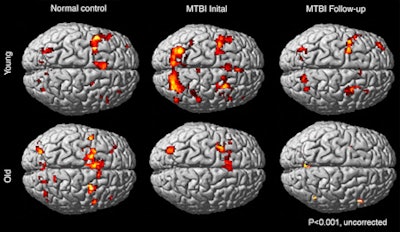 Images show different working memory activation patterns in healthy control subjects and young and old patients with mild TBI. Young control subjects (top left) had increased activation in the frontal and parietal regions, predominantly at the left hemisphere. In the initial exam, young patients with TBI (top middle) had bilateral hyperactivation in the frontal and parietal regions, compared with young control subjects. Partial recovery of the activation pattern (top right) is seen at follow-up imaging. Older control subjects (bottom left) had increased cerebral activation in bilateral frontal and parietal regions. In the initial exam, older TBI patients (bottom middle) showed hypoactivation, compared with the older control subjects. The older TBI patients had even less activation at follow-up (bottom right). Image courtesy of RSNA.
Follow-up scans
At the six-week follow-up, the researchers saw a partial rebound of working memory activation pattern in the younger patients, along with decreased postconcussion symptoms (p = 0.04).
No such changes were detected among the older individuals with mild TBI, which provides "longitudinal functional MR imaging evidence of better neural plasticity in younger patients compared with older patients," the authors wrote.
Based on the results, the influence of age "may lead to future development of separate management strategies for different age groups following mild TBI," Chen and colleagues wrote.
"Functional MR imaging has the potential to provide objective diagnostic and prognostic information in working memory sequelae after mild TBI, as hyperactivation during working memory tasks in younger patients within one month of injury may indicate greater severity of brain injury and neuronal dysfunction," they concluded.


