Advances in technology and software have made 3-tesla MRI a better option for examining fetal abnormalities, with no adverse effects on the fetus, according to a study published in the January issue of the American Journal of Roentgenology.
Researchers from the Children's Hospital of Philadelphia found an "overall advantage" to using 3-tesla MRI with certain imaging sequences to enhance the visualization of structures such as the liver, lungs, kidneys, cartilage, and spine.
The additional information can be critical in identifying the medical needs of the fetus and mother prenatally and postnatally. The increase in signal-to-noise ratio with 3-tesla MRI also could reduce image acquisition time and produce greater spatial resolution (AJR, January 2016, Vol. 206:1, pp. 195-201).
Why MRI?
In the past, 3-tesla MRI has proved its value in pediatric and adult patient populations, but fetal imaging has not benefited from the modality, primarily due to artifacts from the stronger magnet and less-than-adequate image quality, commented lead author Dr. Teresa Victoria, PhD, an assistant professor of radiology at the Perelman School of Medicine at the University of Pennsylvania.
Now, newer-generation MRI scanners and coils and better software have markedly improved the modality, Victoria told AuntMinnie.com.
"From the time we started doing this study three years ago, we were able to get rid of the artifacts, which were impeding the beautiful imaging we now get from fetal MRI," she said.
 Dr. Teresa Victoria, PhD, from the University of Pennsylvania.
The Children's Hospital of Philadelphia is somewhat unique in that it has a dedicated center that cares exclusively for abnormal fetuses.
"When you have an abnormal fetus, you want to make sure you get the right doctor," Victoria said. "We have a group of superb sonographers, but for certain things such as certain lung lesions and gastrointestinal issues, MRI gives you additional details that ultrasound cannot see."
The hospital also prefers MR scans for fetal anomalies when it comes to surgeons and parents.
"An ob/gyn can relate to ultrasound images, but I think the fetal surgeon and parents find it easier to relate and visualize [the anomaly] when they have the MR image," she said. "The clarity of the image makes it easier to come up with a plan of action to save the fetus or do what needs to be done."
To evaluate 3-tesla MRI's efficacy, the researchers retrospectively reviewed 58 fetal MRI studies performed on a 3-tesla scanner (Magnetom Skyra, Siemens Healthcare) between July 2012 and February 2014. They conducted a blind comparison between these cases and 58 scans from a 1.5-tesla system (Magnetom Avanto, Siemens); the fetuses were age-matched and being evaluated for similar abnormalities during the same time period.
The gestational age of the fetuses was 20 to 37 weeks (mean, 30 weeks), and the age range of the mothers was 24 to 44 years (mean, 29 years). Both 3-tesla and 1.5-tesla MRI protocols included single-shot turbo spin-echo (SSTSE), T2-weighted steady-state free precession (SSFP), or true fast imaging with SSFP sequences.
Fetal MRI was performed for a variety of suspected abnormalities, including congenital heart disease, bowel anomalies, lung lesions, congenital diaphragmatic hernia, malformed abdomen (omphalocele), and abdominopelvic mass.
Two experienced pediatric radiologists evaluated the images and rated image quality on a scale of 0 to 4, with 0 indicating that no structure was seen. The scale then progressed from 1 meaning inadequate for diagnostic purposes to 4 indicating excellent image quality and high tissue contrast.
3 tesla over 1.5 tesla
In the overall analysis, one reader had a mean image quality score of 3.1 (± 0.6) for 3-tesla MRI with SSFP, compared with 2 ± 0.5 for 1.5 tesla with SSFP (p < 0.01). The other reader rated 3-tesla MR images with SSFP at 2.0 (± 0.6), compared with 1.8 (± 0.3) for 1.5 tesla with SSFP (p < 0.01).
Collectively, 3-tesla MRI with SSFP sequences achieved higher quality scores than 1.5-tesla MRI with the same sequence for most structures, especially the bowel, liver, lungs, kidneys, cartilage, and spine. Both readers rated 3-tesla MRI as only "marginally better" than 1.5-tesla imaging of the airway.
"The gain is really with the true SSFP," Victoria said. "The depiction of the kidneys is magnificent when you look at it with 3 tesla, and you can see the lungs in much greater detail."
While 3-tesla MRI with SSTSE outperformed 1.5 tesla with SSTSE when imaging the cartilage and spine, the readers found 3-tesla and 1.5-tesla MRI to be statistically comparable and saw little or no difference for the bowel, liver, and airway.
"The lack of an advantage to using 3-tesla MRI with the SSTSE sequence may result from the presence of artifacts seen in association with the use of this sequence, even if the images are still of diagnostic quality," the authors suggested.
Interestingly, image quality scores increased with 1.5-tesla MRI as the fetus aged, while 3-tesla MRI ratings stayed constant regardless of gestational age.
Also, while more imaging artifacts were found with 3-tesla MRI than with 1.5-tesla imaging, their presence "did not limit diagnostic interpretation," the group noted.
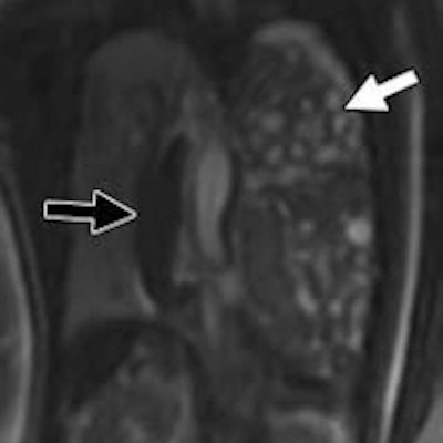
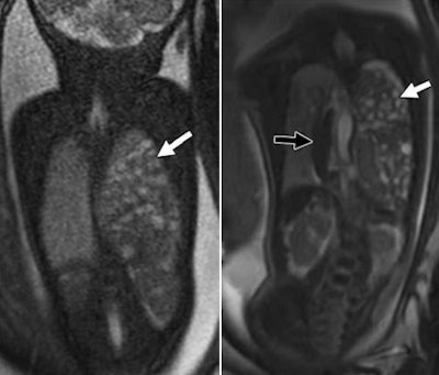
 MR images show two 31-week-old fetuses with left congenital diaphragmatic hernias. Coronal SSFP MR images were obtained at 1.5 tesla (left) and 3 tesla (right). The bowels (white arrows) were herniated in both fetuses, but the bowel wall was better visualized at 3 tesla (right) than at 1.5 tesla (left). A herniated spleen (black arrow, right) was also seen in the 3-tesla image in one fetus. Images courtesy of AJR.
Safety concerns
Prior to the start of this endeavor, the researchers performed their due diligence regarding what effect the doubled magnet strength of 3 tesla might have on a fetus.
One concern was the amount of energy emitted by the MRI scan and absorbed by the fetus. That effect is known as the specific absorption rate (SAR). The U.S. Food and Drug Administration (FDA) mandates that the SAR not exceed 4 W per kg of the mother's body weight for all magnet strengths.
A 2014 study by Victoria et al in Pediatric Radiology concluded that the SAR generated during 3-tesla fetal imaging was well below the FDA limit.
"In any case, all MRI equipment has a built-in system that prohibits exposure beyond FDA limits," Victoria and colleagues wrote in the current paper.
Acoustically, a 3-tesla MRI scan is generally much noisier than its 1-5-tesla counterpart.
"Your concern becomes: What might this do to the fetus?' " Victoria said.
Given that 1.5-tesla MRI has been the standard practice for some 30 years in fetal imaging, indirect data suggest that noise has little effect on the fetus, according to the researchers.
"This is partially because the maternal abdomen provides a protective and isolating layer and also because of the presence of insulating amniotic fluid within the auditory canal," they wrote.
"The last thing you want to do is harm the fetus," Victoria added. "We have done our homework to make sure [3-tesla MRI] is safe, and we will continue to do clinical studies to make sure it is safe."
She also mentioned that contrast agents are never used in fetal imaging, so there are no concerns about the potential harm from such a protocol.
"For fetal imaging, we never give contrast; it is not required for what we do," she said. "It is only possibly required when we do placenta studies, but we don't do those."
Victoria and colleagues plan to continue their efforts to advance 3-tesla MRI for fetal imaging.
"We have to keep working on the artifacts, and make sure we get as clear an image as possible and that the kids are OK," she said. "We are always trying to find better imaging, to improve quality, and to find a better anatomic depiction of whatever you are looking at. This doesn't just happen in the fetal world, but also every field of radiology."


