With the help of functional MRI (fMRI) scans, researchers may have developed a better way to diagnose autism based on anatomic abnormalities in the brain, according to a study published online April 14 in Nature Communications.
Many doctors and scientists think they could improve the diagnosis and understanding of autism spectrum disorders if they had a reliable way to identify specific abnormalities in the brain. Such biomarkers have proven elusive, however, because methods that show promise with one group of patients often fail when applied to another.
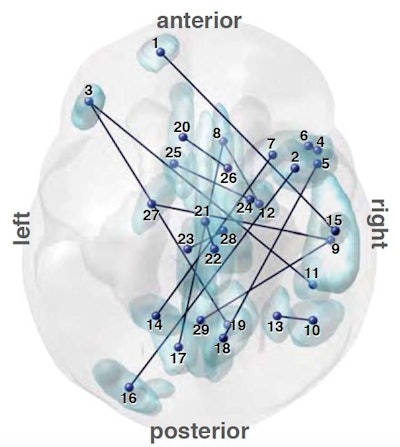
Researchers at the Advanced Telecommunications Research Institute International in Kyoto, Japan, with help from a group at Brown University in Providence, RI, developed a software algorithm that they used to analyze brain network connectivity in Japanese people with and without autism. Based on the analysis, the algorithm found 16 key functional connections between brain regions that enabled it to distinguish the presence of autism with high accuracy between these two sets of people.
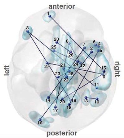 A map of brain connections that proved useful in distinguishing people diagnosed with autism from those without an autism diagnosis. Image courtesy of Brown University.
The algorithm was then applied to a group of 88 adults at seven sites in the U.S. All study volunteers with autism diagnoses had no intellectual disability.
The algorithm averaged 85% accuracy in the Japanese cohort and 75% accuracy among U.S. participants. In another segment of the study, the technique was able to tell the difference between schizophrenia patients, but it could not distinguish between people with depression or individuals with attention deficit/hyperactivity disorder (ADHD).
The researchers noted that subjects only spent about 10 minutes undergoing the fMRI scans, with no need to perform special tasks during the exam.


