The combination of MR arthrography and ultrasound is significantly better than either modality alone for diagnosing medial elbow pain in baseball pitchers who face the prospect of a long recuperation after surgery, according to a study in the June issue of Radiology.
Researchers from Thomas Jefferson University Hospital in Philadelphia found that while MR arthrography and conventional ultrasound individually provide similar diagnoses of ulnar collateral ligament (UCL) tears, using the two modalities together greatly increased sensitivity, specificity, and accuracy (Radiology, June 2016, Vol. 279:3, pp. 827-837).
"You can imagine it is important to have the correct diagnosis before patients undergo surgery, especially in baseball, where there is a lot of money involved and players do not want to undergo surgery if it is not necessary," said lead author Dr. Johannes Roedl, PhD, an attending radiologist in the division of musculoskeletal imaging and interventions.
Elbow stress
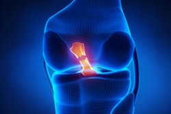
The constant motion of throwing a baseball results in a substantial amount of stress on medial elbow structures, such as the UCL, which stabilizes the medial elbow joint. A tear in this ligament is a common cause of pain among baseball players of all skill levels.
One option for an injured UCL is Tommy John surgery, a procedure named for the first Major League Baseball pitcher to undergo the operation in 1974. Recovery from this surgery can take 10 to 12 months. Even then, two previous studies found that the chance of full rehabilitation and a return to the player's preinjury skill level ranged from 58% to 90%.
 Dr. Johannes Roedl, PhD, from Thomas Jefferson University.
"Tommy John surgery is very common now," Roedl told AuntMinnie.com. "What is scary is that young patients tend to undergo the surgery and it is doubtful that it is always necessary."
While it's been standard practice to use an MR arthrogram with contrast to assess elbow pain, Thomas Jefferson's musculoskeletal ultrasound division about 15 years ago began to explore the concept of adding ultrasound to gather more information.
Musculoskeletal MRI radiologists Dr. Adam Zoga and Dr. William Morrison teamed with musculoskeletal ultrasound radiologist Dr. Levon Nazarian to perform both imaging tests for players from the Philadelphia Phillies baseball team during spring training in Florida. They worked with Phillies' team physician Dr. Michael Ciccotti, an orthopedic surgeon and director of the sports medicine team at Thomas Jefferson University's Rothman Institute. Zoga, Morrison, Nazarian, and Ciccotti are all co-authors of the paper.
The promising results at spring training fueled an interest in adopting the protocol at Thomas Jefferson, where radiologists combined ultrasound with MR arthroscopy to enhance diagnoses of medial elbow pain and suspected UCL tears. With the current study, the researchers for the first time compared the diagnostic accuracy of MRI and ultrasound separately, as well as with the combined approach.
Player profiles
The researchers retrospectively analyzed 144 consecutive male baseball players who presented between January 2003 and January 2013 to a sports medicine clinic for medial elbow pain. The players had no previous surgery and all were older than 15 years of age.
All of the subjects were evaluated with both MR arthrography and stress ultrasound within two days. Surgery and arthroscopy results and clinical follow-up of more than one year were available for all players.
 Co-author Dr. Adam Zoga from Thomas Jefferson University.
The group consisted of 126 pitchers and 18 position players who came from all levels of competition: professional, collegiate, high school, and recreational. They had a mean age of 24.5 years (± 5.6 years) and had injuries to their dominant throwing elbow.
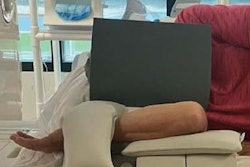
With stress ultrasound, players' elbows were flexed at approximately 30° and held in that position by one of Thomas Jefferson's resident physicians. For rest ultrasound, the elbow remained at that angle without valgus stress and the width -- or gap -- of the ulnotrochlear joint space at the UCL was measured on the ultrasound screen. Increased joint gapping indicates a tear of the UCL.
"The ligament essentially provides stability. If it tears or is injured, you can 'gap open' the joint when there is stress on [the UCL]," Roedl explained. "We wanted to look at what measurement should be used? What is the threshold? What is too much of an opening that would indicate a tear?"
All elbow MR arthrograms were performed on 1.5-tesla scanners (Signa or Optima, GE Healthcare) after intra-articular injection of gadopentetate dimeglumine contrast (Magnevist, Bayer HealthCare Pharmaceuticals) with fluoroscopy.
Surgery or arthroscopy was performed on all 144 patients within three weeks of the MR and ultrasound examination. Results were used as the gold standard to evaluate the diagnostic accuracy of MRI and ultrasound individually and collectively.
Modality comparisons
Among the 144 baseball players, 53 had UCL tears, 31 had nerve inflammation (ulnar neuritis), 59 had inflammation of the tendon and muscle (myotenositis), and 48 had inflammation of the bone and its overlying cartilage (osteochondritis).
When they examined the images of each player's elbows, the researchers found a significant difference in the joint gap of the injured elbow (3.7 mm ± 1.3 mm) versus the joint gap of the contralateral, or uninjured, elbow (1 mm ± 0.3 mm). Using a joint gap threshold of greater than 1 mm between the injured and uninjured elbows, the combination of ultrasound and MR produced greater sensitivity, specificity, and accuracy for diagnosing UCL tears.
| Comparison of US and MR arthrography for UCL tears | |||
| Stress ultrasound | MR arthrography | Ultrasound + MR | |
| Sensitivity | 96% | 81% | 96% |
| Specificity | 81% | 91% | 99% |
| Accuracy | 87% | 88% | 98% |
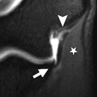
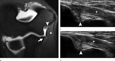 Images are of a 21-year-old baseball pitcher with a full-thickness UCL tear. MR arthrography (a) and corresponding medial elbow ultrasound exams at rest (b) and with stress (c). Full-thickness tear at the proximal UCL (arrowhead) and additional small partial-thickness tear at the distal UCL (arrow). Normal common flexor muscle bulk (star) is also shown. Substantial gapping (5.7 mm) at the ulnotrochlear joint between rest (rest gap: 2.0 mm, white line in b) and stress (stress gap: 7.7 mm, white line in c) is illustrated. Image courtesy of Radiology.
For the 31 patients with ulnar neuritis, MR arthrography alone posted a sensitivity of 74%, specificity of 92%, and accuracy of 88%. When combined with ultrasound, sensitivity rose to 90%, specificity to 100%, and accuracy to 98%.
Interestingly, for the cases of myotenositis and osteochondritis, MR arthroscopy was significantly superior to ultrasound, which added no diagnostic value.
"When it comes to other injuries that baseball pitchers have at the medial elbow, MRI is usually better, especially injuries to the bone," Roedl said. "Ultrasound is not good for bone. If someone has a bone bruise or anything dealing with cartilage inside the joint, you need the MRI."
Of course, with better diagnoses come more appropriate treatment and therapy regimens for injured players, especially for younger athletes.
"There are many ambitious parents out there who jump into surgery when their kid has medial elbow pain," Roedl said. "So [Tommy John surgery] is probably overperformed at this point, which is why this is such an important imaging test and study."
He added that Thomas Jefferson plans to expand the clinical use of both MR arthrography and ultrasound, especially for the "weekend warriors" who are injured playing baseball.
"It is very important to get the message across that although the study results were very good for ultrasound, if I had to pick just one test, I would still go with the MRI, because you can't really see the bones and cartilage with ultrasound at all," Roedl emphasized. "That is an important take-home message."


