By taking advantage of parallel imaging on the latest generation of 1.5- and 3-tesla scanners, researchers have devised a quick five-minute shoulder MRI protocol that still produces diagnostic-quality images, according to a study in the April issue of the American Journal of Roentgenology.
The new protocol reduced average scan times by 62% to 72%, depending on the scanner's magnet strength, and still afforded readers the ability to detect major abnormalities at a rate similar to using images from standard 2D fast spin-echo (FSE) sequences.
 Dr. Naveen Subhas from the Cleveland Clinic.
"These techniques will be most useful in patients who have a difficult time tolerating an MRI scan because of claustrophobia or discomfort," said lead author Dr. Naveen Subhas, an associate professor of radiology at the Cleveland Clinic. "The key is to be able to do parallel imaging, which is how we accelerate the protocol."
Time and efficiency
MRI is considered the modality of choice to evaluate shoulder conditions such as rotator cuff tears, labral abnormalities, and other causes of shoulder pain. The downside of MRI is that cost and scanner availability can limit its use, the authors wrote (AJR, April 2017, Vol. 208:4, pp. W146-W154).
The Cleveland Clinic allots 30- to 40-minute time slots for MRI scans, with actual imaging time accounting for approximately 15 to 20 minutes, Subhas said. The rest of the time is spent positioning the patient.
"We figured that if we really wanted to be more efficient, cutting the scan time from 15 minutes to 10 minutes can make a big difference," he added.
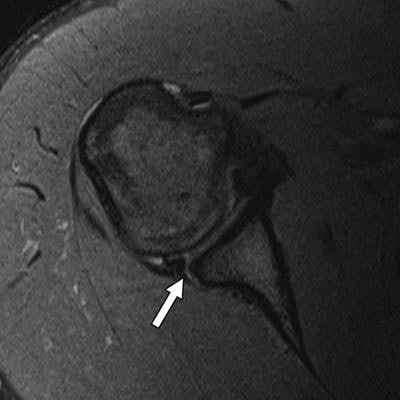
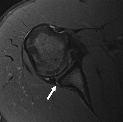
 A posterior labral tear (arrow) in a 20-year-old man detected with axial T2-weighted multiplanar 2D fast spin-echo unenhanced standard MRI (above) and the fast five-minute protocol (below) using 3-tesla MRI. Images courtesy of AJR.
The key component to reducing imaging time and maintaining diagnostic image quality is having a scanner that can perform parallel imaging. The technology shortens exam time by obtaining a signal from a reduced field-of-view with fewer phase-encoding steps, which allows "images to be acquired with the same resolution as that obtained with a standard sequence in less time," the authors wrote.
"Not only does the scanner have to do parallel imaging, but the coil used to image the body part also must be compatible with parallel imaging," Subhas said.
The current retrospective study included MRI scans obtained at two academic imaging centers between December 2014 and May 2015. Three musculoskeletal radiologists reviewed 151 shoulder exams performed on a 3-tesla scanner (Magnetom Verio, Siemens Healthineers) with a 4-channel shoulder array coil, as well as 50 shoulder exams performed on a 1.5-tesla system (Magnetom Aera, Siemens) with a 16-channel shoulder array coil.
On the clock
To compare scan times and subsequent image quality, standard 2D FSE shoulder MR sequences were immediately followed by the fast five-minute parallel imaging technique. Among the subjects, 66 were being scanned for unknown shoulder pain, 49 for rotator cuff tears, and 22 for labral tears or biceps tendon abnormality.
| Length of scanning time by MRI protocol | |||
| MRI strength | Standard MRI protocol | Fast MRI protocol | Difference |
| 3 tesla | 14 minutes, 6 seconds | 5 minutes, 23 seconds | 62% |
| 1.5 tesla | 15 minutes, 39 seconds | 4 minutes, 30 seconds | 72% |
Both imaging techniques achieved similar rates of detection for major abnormalities. For example, 102 (75%) of 135 full-thickness shoulder tears and 32 (68%) of 47 complete biceps tendon tears were detected by both protocols (p ≥ 0.08 for each structure).
"In other words, readers were not more likely to report major findings on standard MRI compared with fast MRI," the authors wrote.
In addition, sensitivity for the five-minute MRI protocol was equal to or better than sensitivity for the standard MRI technique for every structure except glenohumeral cartilage defects, which were limited by a small sample size on the 1.5-tesla scanner, the researchers noted.
The right tools
Now that the results have been published, Subhas and colleagues plan to use the five-minute MRI protocol in the clinical setting for patients with issues such as claustrophobia or anxiety during scans.
To what degree could the five-minute MRI protocol spread beyond musculoskeletal imaging? Subhas speculated that brain imaging could be one possibility, as long as the shortened MRI protocol does not detract from achieving an accurate diagnosis.
"Obviously one of the compromises we make with this technique is that our ability to see fine detail is somewhat compromised," he said. "The question is: How much fine detail do we need to see? That depends on what we are imaging. [The fast MRI protocol] is certainly doable in other body parts, but we will have to see if the results will be the same."


