Thursday, November 30 | 11:20 a.m.-11:30 a.m. | SSQ13-06 | Room E451A
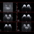
Researchers from Johns Hopkins University have developed a novel MRI technique that achieves high diagnostic accuracy in the surveillance of patients following resection and reconstruction with a tumor prosthesis.The protocol is called sequences for metal artifact reduction in tumor and trauma (SMART), and it is designed to improve visualization around metal objects.
"Two types of sequences are employed: those that reduce artifact adjacent to the metal, and those that improve visualization of the tissues that are more remote from the site of the metal," explained Dr. Laura Fayad, chief of musculoskeletal imaging and director of the radiology department's translational research program.
In the study to be presented at RSNA 2017, 15 subjects with tumor prostheses underwent 17 1.5-tesla MRI scans. The quality of each SMART sequence adjacent to and remote from the prosthesis was rated on a scale of 1 to 4, with 1 meaning "greater than 75% artifact" to 4 meaning "no substantial artifact."
Among the findings, a receiver operating characteristic (ROC) analysis showed improved diagnostic performance using the successive addition of fast slice encoding for metal artifact correction, dynamic contrast-enhanced MRI, and conventional metal reduction.
The ability to use MRI to evaluate prostheses could be a "great advantage" over other cross-sectional modalities because MRI generally offers "excellent contrast resolution" for detecting both bone marrow and soft-tissue abnormalities, Fayad said.
"In the case of a tumor prosthesis placed following surgical resection of a sarcoma, the detection of a recurrent tumor was traditionally hampered by the presence of metal in the surgical bed and field-of-view," she said. "Now, with SMART imaging, we are able to detect recurrences as well as complications around tumor prostheses with great accuracy, hence offering a significant advance in patient care."


.fFmgij6Hin.png?auto=compress%2Cformat&fit=crop&h=100&q=70&w=100)


.fFmgij6Hin.png?auto=compress%2Cformat&fit=crop&h=167&q=70&w=250)


