Recovering from weightlessness might be the least of the problems that astronauts face when they return home to Earth. MRI scans revealed a loss of gray-matter volume in the brain soon after touchdown and prolonged changes in circulation of cerebrospinal fluid (CSF) some seven months later, according to a study published in the October 25 issue of the New England Journal of Medicine.
The good news is that gray-matter volume returned close to preflight levels in less than a year. White-matter brain tissue volume appeared unchanged on MRI immediately after landing. However, seven months later, scans displayed a widespread reduction in white-matter volume. The researchers speculated that over that time, the volume of the white matter may slowly be replaced by an influx of CSF (NEJM, October 25, 2018, Vol. 379:17, pp. 1678-1680).
"Whether or not the extensive alterations shown in the gray and the white matter lead to any changes in cognition remains unclear at present," said study co-author Dr. Peter zu Eulenburg, a professor at Ludwig Maximilian University in Munich, in a statement. "So far the only clinical indication for detrimental effects is a reduction in visual acuity that was demonstrated in several long-term space travelers."
These changes may very well be attributable to the increased pressure exerted by the cerebrospinal fluid on the retina and the optic nerve.
Previous research has shown that extended spaceflight can adversely affect brain anatomy, including the narrowing of CSF spaces, a decrease in frontotemporal volume, and an increase in ventricle size.
"However, information is limited about the effects of microgravity on brain volume, particularly regarding changes that are evident more than one month after spaceflight," the authors noted.
In this prospective study, the researchers collected data from T1-weighted MRI scans performed on 10 male astronauts (mean age, 44 years) who spent an average of 189 days on the International Space Station. Scans were conducted prior to their travel and an average of nine days after their return to Earth. In addition, seven men were scanned again seven months after their return to Earth.
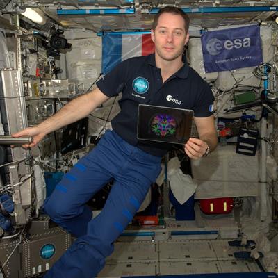
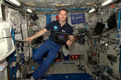
 French astronaut Thomas Pasquet shows an MR image of his brain taken before he boarded the International Space Station. He returned to Earth in June 2017 after spending 196 days in orbit. Image courtesy of the European Space Agency and NASA.
The first MRI scan upon landing revealed a decrease in gray-matter volume in the orbitofrontal and temporal cortexes of the astronauts, compared with preflight scans. The greatest loss (3%) was in the right middle temporal gyrus. Interestingly, by the second postflight scan seven months later, gray-matter volume almost rebounded to preflight levels, with a lingering reduction of 1.2% in the right temporal gyrus.
The first follow-up MRI scan also showed increased CSF volume within the cortex, compared with preflight results, with the greatest increase (13%) in the third ventricle. The researchers also suggested that increased pressure exerted by CSF on the retina and the optic nerve might be the cause for the astronauts' vision problems.
At this point, it is too early to know how these findings someday might influence more earthly clinical applications.
"More research into the cognitive and neurological consequences of our findings is needed," zu Eulenburg wrote in an email to AuntMinnie.com. "More data was acquired and will be published over the next year."


