Thursday, November 29 | 11:30 a.m.-11:40 a.m. | RC613-14 | Room S502AB
With the continuing controversy over gadolinium retention in bone, skin, liver, and portions of the brain long after contrast-enhanced MRI scans occur, exploration continues for viable alternatives, as researchers will discuss in this presentation.One possibility is ferumoxytol (Feraheme, Amag Pharmaceuticals), which contains iron oxide nanoparticles that can be detected with MRI. At this point, however, it is unknown whether ferumoxytol leaves residue in the brain, similar to the effects of gadolinium.
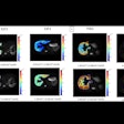
Dr. Ashok Joseph Theruvath, a postdoctoral research fellow at Stanford University School of Medicine, is scheduled to present a retrospective case-control study of ferumoxytol. The study included 12 pediatric patients who received at least two intravenous injections of ferumoxytol for 3-tesla brain MRI scans and 12 additional patients who served as unexposed controls. The researchers recorded visual susceptibility effects and R2* values of the caudate, globus pallidus, putamen, dentate nucleus, thalamus, and substantia nigra.
They found no significant difference in visual susceptibility effects and quantitative R2* data for all brain regions. However, follow-up studies some 15 months later revealed slightly increased R2* in the dentate nucleus and globus pallidus, which suggests that ferumoxytol "might be transiently retained in specific brain regions," according to Theruvath and colleagues.
"Further studies have to evaluate potential minimal and transient retention of iron oxides in the brain," they wrote.


.fFmgij6Hin.png?auto=compress%2Cformat&fit=crop&h=100&q=70&w=100)


.fFmgij6Hin.png?auto=compress%2Cformat&fit=crop&h=167&q=70&w=250)


