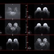
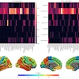
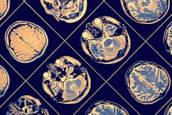
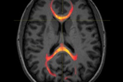
By combining structural and functional MRI (fMRI), Canadian and Dutch researchers have identified three unique patterns in the brain to better monitor concussions in young female athletes, according to a study published online December 3 in NeuroImage: Clinical.
Using 3-tesla MR imaging, the researchers were able to identify acute brain changes after an athlete has experienced a concussion, persistent brain changes six months after the injury, and evidence of an athlete's concussion history.
The study included 52 female athletes from the women's varsity rugby team at Western University in London, Ontario. Among the subjects, 21 had a concussion during one regular season of play. The researchers used a technique that combines multiple imaging measures to simultaneously look at structural and functional information.
The presence of structural and functional brain changes six months after the injury supports prior research showing persistent changes, according to the group.
"This study highlights the important contributions advanced imaging techniques can make in helping clinicians understand what happens biologically in the brain when players become concussed -- and these improvements translate into better clinical decisions," said Christian Beckmann, PhD, from Radboud University Medical Centre Nijmegen, in a statement from Western University.


.fFmgij6Hin.png?auto=compress%2Cformat&fit=crop&h=100&q=70&w=100)

.fFmgij6Hin.png?auto=compress%2Cformat&fit=crop&h=167&q=70&w=250)


