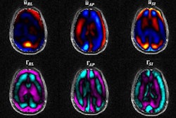
U.S. and European researchers could be on the brink of reducing the time of an MRI brain scan to milliseconds with an MR elastography technique, according to a preclinical study published online April 17 in Science Advances.
Their approach uses functional MR elastography (fMRE) to create maps of tissue stiffness, which in turn track the activity of brain function that occurs in bursts as short as 100 msec. Early indications are that fMRE could be better than functional MRI (fMRI), since fMRI cannot keep pace with the split-second activity of the brain's neurons as they process thoughts and react to various stimuli.
"Traditional fMRI has a temporal response of several seconds and, therefore, cannot measure high-level cognitive processes that evolve in tens of milliseconds," wrote author Samuel Patz, PhD, from Brigham and Women's Hospital in Boston, along with colleagues from King's College London in the U.K. and the French National Institute for Health and Medical Research (INSERM) in Paris. "To advance neuroscience, imaging of fast neuronal processes is required. Here, we directly show in vivo imaging of fast neuronal processes at 100-ms time scales by quantifying brain biomechanics noninvasively with MR elastography."
The researchers explain their new approach. Video courtesy of Patz et al.
This five-year collaboration took a rather unusual turn after an unexpected discovery. Researchers were exploring MR elastography for lung imaging when they tried the modality on mouse brains. The images inexplicably revealed a stiffening of approximately 10% in the somatosensory cortex of rodents after repeated electric stimulation with frequencies ranging from 0.1 Hz to 10 Hz.
Patz and colleagues followed up with another fMRE scan. This time they chose to plug the mouse's ear canals with a gel to reduce possible stimulation of the auditory cortex from preclinical MRI scanner noise. They subsequently observed a softening of the mouse's auditory cortex on the side of the brain that processed sound from the plugged ear.
The brain's stiffening and softening responses were replicated on subsequent fMRE scans under different types and levels of stimuli. At 100 msec of stimulation, there also were changes in the thalamus, which is "the relay location [for] input to the cortex," the authors noted.
"We anticipate that mapping neuronal activity by the measurement of tissue stiffness will provide a new methodology for studying brain function at high temporal and spatial resolution with specific application to tracking neural circuitry at high speed," they added. "We look forward to future studies of neuronal propagation with different stimuli at different stimulus switching frequencies, which will allow for the elucidation of the different neuromechanical coupling mechanisms with their different amplitudes and time constants."
Naturally, Patz and colleagues want to advance this fMRE protocol to observe neuronal activity in humans. The potential benefits include better understanding and diagnoses of neurological pathologies in which neuronal activity is abnormal or dysfunctional.
"The translation of our approach to humans is imminent, and we have already obtained preliminary data in the human visual cortex," the authors wrote. "Thus, fMRE has great potential to facilitate and deepen the understanding of the pathway of neuronal signals propagating in the in vivo brain, including the elucidation of impaired neural circuitry associated with pathologic subcomponents of neuronal physiology."


