MONTREAL - While 7-tesla MRI offers high resolution and strong contrast, its shortcoming is the lack of field homogeneity. This can be addressed with the use of dielectric pads, according to poster research presented at the Society for MR Radiographers & Technologists (SMRT) annual meeting.
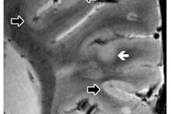
"The 7T [scanner] gives you better quality images," said Huijun Liao, an MRI technologist at Brigham and Women's Hospital in Boston. "We use it for research and clinical purposes. We saw, however, that there has been severe signal drop-off, which affected diagnosis. If you see dark spots here and there, how do you know if there is an intact cerebellum or intact occipital lobe?"
New technologies like 7-tesla MRI present challenges to technologists who seek to optimize image quality, Liao said. When the brain is imaged to obtain visualization of a tumor, for example, there may be a reduced signal-to-noise ratio where the tumor is located, she explained.
"How do you see how big the tumor is if there is signal drop-off at the tumor area?" Liao asked the audience. "We need a quick, easy solution that would solve the homogeneity problem for a fast-paced clinical environment."
One way to overcome the homogeneity issue is the placement of dielectric pads, which contain conductive material and are designed to reduce dielectric artifacts. The pads are placed between the patient and the receiver coil.
Liao and colleagues decided to investigate the impact of the pads on brain imaging. She noted that previous research has shown that dielectric pads effectively increase image quality.
"We wanted to see if there is a benefit to [using dielectric pads in] imaging and how homogeneous it will be," she said. "We want it to be homogeneous, so we get a good image and good signal-to-noise ratio."
Liao and co-investigators performed MRI exams on a 7-tesla MRI scanner with a commercially available head coil (Nova Medical). When performing the scan, the researchers placed two thin dielectric pads on both sides of a subject's head. The scan was performed on a subject with and without pads to assess the effects of dielectric pads, with scans performed of the posterior cingulate gyrus, the left superior temporal gyrus, and the left frontal lobe white matter -- including caudate nucleus in phantoms, control subjects, and subjects with schizophrenia.
In terms of results, homogeneity at full width half maximum was enhanced in all three regions in a healthy control scan, and the posterior cingulate gyrus -- one of the most homogenous areas of the brain and an area that is typically involved in MR spectroscopy studies -- demonstrated the largest improvement, Liao said.
The mean signal-to-noise ratio with the pads was 453 in six subjects, while it was 316 in 10 subjects without use of the pads, she pointed out.
The signal-to-noise ratio, however, was only improved in one area of the brain when dielectric pads were used: the left superior temporal gyrus. This indicated significant benefit for MR spectroscopy voxels in brain regions proximal to dielectric pads, Liao explained. The signal-to-noise ratio was 43% higher, deemed a significant increase in that area -- a region of the brain regarded as critical for patients with schizophrenia and other mental disorders.
One of the challenges with the placement of dielectric pads has been their use in patients who have larger head circumferences, Liao noted. Because the sizes of available head coils are often limited, investigators have had to modify the placement of the pads in individuals with bigger head circumferences. In these clinical cases, technologists will place at least one pad on the back of the head to ensure image quality, Liao added.


