Findings from MRI scans show that vaping immediately affects vascular function, even when the liquid solution contained in the electronic cigarette (e-cigarette) does not contain nicotine, according to a study published August 20 in Radiology.
The results demonstrate that, even without nicotine, e-cigarettes are harmful to the body, said lead author Alessandra Caporale, PhD, of the University of Pennsylvania in Philadelphia, in a statement released by the RSNA.
"These products are advertised as not harmful, and many e-cigarette users are convinced that they are just inhaling water vapor," Caporale said. "But the solvents, flavorings, and additives in the liquid base, after vaporization, expose users to multiple insults to the respiratory tract and blood vessels."
E-cigarettes deliver nicotine by the electric heating and aerosolization of a flavored solution, Caporale and colleagues wrote. More than 9 million U.S. adults vape, according to the researchers, who cited data from the U.S. Centers for Disease Control and Prevention. More disturbing is the fact that using e-cigarettes has become prevalent among teenagers: In fact, last year's National Youth Tobacco Survey findings show that in 2018 more than 3.6 million middle and high school students were vaping.
The liquid solution in e-cigarettes contains potentially harmful toxic substances, and, when inhaled, these substances enter the alveoli of the lungs, are absorbed by blood vessels, and can interfere with vascular function, Caporale and colleagues wrote.
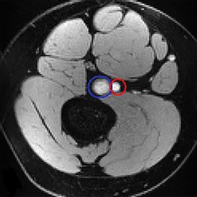
The researchers sought to investigate the effects of vaping on systemic vascular function by conducting a study that included 31 healthy nonsmoking adults (mean age, 24) who underwent MRI scans before and after vaping with an e-cigarette that did not contain nicotine. The team used MRI to scan the femoral artery in participants' legs, as well as their aortas and brains. For the femoral artery exam, the researchers constricted and then released participants' blood flow in the upper leg using a cuff; the brain MRI imaged the sagittal sinus during a series of 30-second breath-holds and normal breathing.
Caporale and colleagues found that a single vaping episode cut blood flow and weakened vascular reactivity in the femoral artery, with a 34% reduction in flow-mediated dilation (the dilation of an artery mediated by blood flow increase), a 17.5% reduction in peak flow, and a 25.8% reduction in blood acceleration.
As for the aorta and the brain, the group found that vaping caused a 3% increase in aortic pulse wave velocity -- which suggests aortic stiffening -- but no statistically significant changes in cerebrovascular reactivity.
![Response to cuff occlusion in the femoral circulation. A: Vessel-wall images of the superficial femoral artery (SFA) at different points (60 secs [A60], 90 secs [A90], and 120 secs [A120]) as indicated by crosses in B during reactive hyperemia. The dashed circles represent the lumen area at baseline. B: Superficial femoral vein (SFV) oxygen saturation (SvO2) at baseline (green line) and during hyperemia. C: SFA blood flow velocity (V). D: Axial image obtained with MRI in the thigh, with SFA and SFV indicated in red and blue, respectively. Δ SvO2 = peak-to-peak SvO2, HI = hyperemic index, PFR = peripheral flow reserve, RI = resistivity index, TFF = time of forward flow, TP = time to peak, TW = washout time, Vb = baseline velocity, VP = peak hyperemic velocity, Vr = retrograde velocity during early diastole, Vs = systolic velocity. Image courtesy of the RSNA.](https://img.auntminnie.com/files/base/smg/all/image/2019/08/am.2019_08_20_18_35_1507_2019_08_20_vaping_MRI.png?auto=format%2Ccompress&fit=max&q=70&w=400) Response to cuff occlusion in the femoral circulation. A: Vessel-wall images of the superficial femoral artery (SFA) at different points (60 secs [A60], 90 secs [A90], and 120 secs [A120]) as indicated by crosses in B during reactive hyperemia. The dashed circles represent the lumen area at baseline. B: Superficial femoral vein (SFV) oxygen saturation (SvO2) at baseline (green line) and during hyperemia. C: SFA blood flow velocity (V). D: Axial image obtained with MRI in the thigh, with SFA and SFV indicated in red and blue, respectively. Δ SvO2 = peak-to-peak SvO2, HI = hyperemic index, PFR = peripheral flow reserve, RI = resistivity index, TFF = time of forward flow, TP = time to peak, TW = washout time, Vb = baseline velocity, VP = peak hyperemic velocity, Vr = retrograde velocity during early diastole, Vs = systolic velocity. Image courtesy of the RSNA.
Response to cuff occlusion in the femoral circulation. A: Vessel-wall images of the superficial femoral artery (SFA) at different points (60 secs [A60], 90 secs [A90], and 120 secs [A120]) as indicated by crosses in B during reactive hyperemia. The dashed circles represent the lumen area at baseline. B: Superficial femoral vein (SFV) oxygen saturation (SvO2) at baseline (green line) and during hyperemia. C: SFA blood flow velocity (V). D: Axial image obtained with MRI in the thigh, with SFA and SFV indicated in red and blue, respectively. Δ SvO2 = peak-to-peak SvO2, HI = hyperemic index, PFR = peripheral flow reserve, RI = resistivity index, TFF = time of forward flow, TP = time to peak, TW = washout time, Vb = baseline velocity, VP = peak hyperemic velocity, Vr = retrograde velocity during early diastole, Vs = systolic velocity. Image courtesy of the RSNA.These findings suggest that vaping impairs the function of the endothelium. However, the researchers noted that more research is needed.
"Our study provides further insight into the effects on the endothelium from electronic cigarette exhalants detectable by noninvasive quantitative MRI markers," the group concluded. "In light of these results, it would be desirable to corroborate our findings in larger cohorts."


