Thursday, December 5 | 11:30 a.m.-11:40 a.m. | SSQ09-07 | Room E351
When fetuses do not grow and develop adequately in the womb, clinicians and obstetricians can turn to MRI for a noninvasive view of the problem, according to this Thursday morning presentation.The issue is often a condition known as intrauterine growth restriction (IUGR), in which a fetus does not grow at a normal rate and is smaller than his or her gestational age. The stunted growth can lead to poor health and related defects after birth that slow a baby's overall development.
To investigate the effects of IUGR, a team led by Dr. Behnaz Moradi, an assistant professor of radiology at Tehran University of Medical Sciences in Iran, performed 3-tesla MRI scans on 42 pregnant women with IUGR and 28 age-matched pregnant women with normally growing fetuses. There were no significant differences in maternal characteristics and fetal gestational age between the two groups.
The MRI protocol included T2-weighted half-Fourier acquisition single-shot turbo spin echo (HASTE) to assess several regions of the brain. The researchers specifically targeted cortical thickness in the insula, frontal, occipital, and temporal regions. They also measured whole brain area and the area of the frontal, temporal, occipital, cerebellum, midbrain, and pons regions. Any cases with brain structural anomalies were excluded, and the development of all fetuses was monitored until birth.
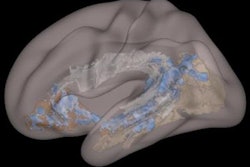
Moradi and colleagues found significantly lower birth weight among the fetuses with intrauterine growth restriction. While brain signal was normal in all cases, they observed significantly thinner cortex in the insula and temporal lobes of IUGR fetuses, compared with the control group, as well as significantly smaller whole brain area.
MRI has the potential to add valuable clinical information regarding the development of fetuses with intrauterine growth restriction, they concluded.


