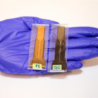
Researchers at San Diego State University (SDSU) are developing a glassy carbon electrode for use with MRI scans during deep brain stimulation, according to a study published online November 18 in Nature Microsystems and Nanoengineering.
The carbon device, codeveloped with the Karlsruhe Institute of Technology (KIT) in Germany, is designed to emit and receive string signals for a long time without corroding or deteriorating. The researchers last year showed that their carbon electrode survived 3.5 billion cycles of electrical impulses, compared with 100 million cycles of electrical impulses with a metal electrode.
Deep brain stimulation uses electrodes implanted in a person's brain to produce electrical impulses to control abnormal movement. The procedure is used for patients with movement disorders -- such as Parkinson's disease, tremors, and uncontrolled muscle contractions known as dystonia -- who do not respond to medication.
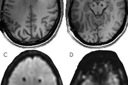
Current metal-based electrodes can produce heat and create image artifacts in MRI scans, thus blocking views of abnormalities in the brain. They also can become magnetized from the bore's magnet vibrations when patients undergo MRI scans.
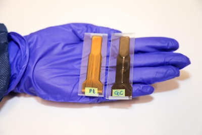 A side-by-side look at platinum (left) and glassy carbon (right) thin-film electrodes for deep brain stimulation. Image courtesy of San Diego State University.
SDSU and KIT researchers tested the carbon electrodes directly in an MRI scanner and confirmed that they did not become magnetized and did not irritate the patient's brain. They plan to develop the technology further and continue MRI safety tests.


