Researchers from the U.K. have retooled their prototype magnetoencephalography (MEG) scanner to be able to capture 3D brain images of active adults and children. The new device may improve current understanding of brain development and neurological conditions, according to an article recently published online in Nature Communications.
"We designed and built a new [MEG] bike helmet-style design, and using this we were able to successfully analyze brain activity of a 2- and 5-year-old whilst they were doing an everyday activity," said lead author Ryan Hill, a doctoral student at the University of Nottingham, in a university statement.
The revamped MEG scanner provides researchers with the unprecedented opportunity to study a wide range of neurological and mental health conditions, such as epilepsy and autism, in children based on their brain activity, Hill and colleagues noted.
A pediatric MEG scanner
For years, various groups have studied brain structure and function using MRI and wearable technologies such as MEG and electroencephalography. Recent developments in quantum technology have bolstered this work by allowing researchers to measure brain activity from the pattern of magnetic fields generated outside the head using optically pumped magnetometers (OPMs).
In a prior proof-of-principle study, Hill and colleagues presented a prototype MEG scanner as part of a five-year project to advance research and treatment for patients with neurodegenerative disorders. The scanner consisted of OPMs attached to a large, individually tailored 3D-printed helmet that was capable of mapping brain function in adults as they performed basic tasks.
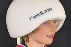
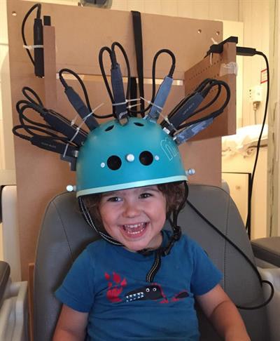 A child undergoing imaging with the MEG helmet scanner. Image courtesy of Dr. Rebeccah Slater from the University of Oxford.
The prototype MEG scanner, however, provided limited information on early brain development due to most infants' inability to tolerate the imaging environment and inherent design restrictions preventing natural movement during imaging.
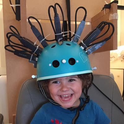
Seeking to resolve these issues, the researchers redesigned their MEG scanner to include lightweight OPMs mounted atop a generic bike helmet that could fit the head of most children, including infants as young as 2 years old (Nat Commun, November 5, 2019, Vol. 10:4785).
The new scanner weighs roughly 0.88 lb (400 g) and holds an infrared camera and electromagnetic coils that can nullify the background magnetic field. The joint camera and coil system allows wearers to move their head freely during scanning.
"The initial prototype scanner was a 3D-printed helmet that was bespoke -- in other words, only one person could use it," Hill said. "It was very heavy and quite scary to look at. Here, we wanted to adapt it for use with children, which meant we had to design something much lighter and more comfortable but that still allowed good enough contact with the quantum sensors to pick up signals from the brain."
Mapping brain activity
The researchers first tested the helmet's capacity to record brain activity in a 2-year-old and a 5-year-old. Both children underwent scanning as their mother gently touched the back of their hands. A 24-year-old woman also underwent scanning under similar conditions.
The team found that touch stimulation triggered clear changes in brain activity, which they measured using synthesized gradiometers.
Subsequently generating 3D images of the brain activity was a multistep process that involved acquiring a CT scan of the helmet fitted with sensors, converting the scans in virtual 3D models, and using a 3D digitizer to capture the shape of the participants' head and face.
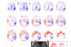
The researchers used these images, as well as data from a motion-tracking camera, to coregister the location and orientation of the helmet OPMs relative to the participants' head. This technique ultimately allowed them to generate 3D images representing changes to beta brainwave activity.
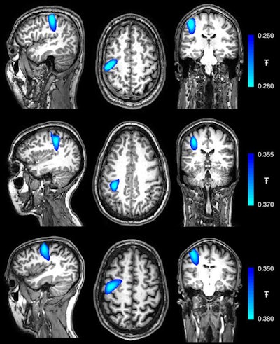 Coregistereed images showing the spatial signature of beta modulation; the largest peak was found in precentral-sulcus (motor cortex) in all three subjects. Image courtesy of Hill et al. Licensed under CC BY-NC 4.0.
Following the early tests, Hill and colleagues used their MEG scanner to examine the brain activity of a 14-year-old teenager while completing a simple, repetitive task on a computer and of a 24-year-old woman as she played a ukulele.
Despite the substantial head movement that these tasks required, the MEG scanner was able to record clear electrophysiological responses in the brain. Of note, the largest change in brain activity while the participants performed their respective tasks occurred in the contralateral primary somatosensory cortex of the brain.
The ability to examine brain activity while an individual engages in tasks naturally was previously unthinkable with conventional brain imaging equipment, the authors noted. The new MEG helmet design may now pave the way for neurodevelopmental research at most ages.
"This study is a hugely important step toward getting MEG closer to being used in a clinical setting, showing it has real potential for use in children," senior author Matthew Brookes, PhD, said. "The challenge now is to expand this further, realizing the theoretical benefits, such as high sensitivity and spatial resolution, and refining the system design and fabrication, taking it away from the laboratory and toward a commercial product."

