The claims of companies are alluring: "Enlarge and reshape the buttocks and add shape to the hips. Fillers are an easier way to achieve the buttock shape you want, without surgery." But the adverts don't tell customers that the long-term results can be disastrous, and more clinicians and radiologists are seeing patients in need of urgent help, according to award-winning research from Latin America.
"Silicone can generate multiple complications, regardless of the injection site. The true incidence and prevalence are unknown, although its frequency is increasing and it has become a public health problem to which we must pay greater attention," said Dr. John Jairo Echeverri, a radiologist at Imbanaco Medical Center in Cali, Colombia.
The use of silicone for cosmetic purposes persists worldwide, and it appears to be growing fast in developing countries, despite the ban on silicone for cosmetic use by the U.S. Food and Drug Administration and other regulatory agencies. Radiologists must take them into account and learn to recognize the images that they generate, and it is essential to consider that silicone can later migrate to other locations beyond the injection site.
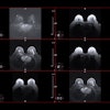
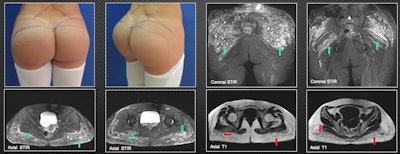 A 29-year-old patient was injected with biopolymers in the gluteal region six years ago to increase volume. She is currently asymptomatic, but she decided to schedule surgical removal of the material. MRI shows multiple nodular and other elongated lesions in the subcutaneous cellular tissue of the gluteal region. They are hypointense on T1-weighted images (red arrows) and hyperintense on T2-weighted and short-tau inversion recovery images (green arrows). This is compatible with biopolymers, which also compromise the maximum gluteal muscles bilaterally, especially on the left side; the other muscle groups in the pelvis show no infiltration by biopolymers. All figures courtesy of Dr. John J. Echeverri and RSNA 2020.
Awareness of iatrogenic allogenosis, or foreign modeling agent reaction, must increase because it has a high socioeconomic impact and is not covered well in the literature, Echeverri and colleagues explained in an RSNA 2020 e-poster for which they received a certificate of merit. Get to know the multiple early and late complications and the different imaging modalities used in assessment of patients, they recommended.
Initially, scientists thought silicone was inert, but its liquid form can produce adverse reactions such as inflammation, induration, ulceration, migration, and granuloma formation. Iatrogenic allogenosis is now thought to affect more than one million people in Latin America.
Biopolymers are dangerous because the organism identifies the biopolymer as a foreign body and triggers an excessive inflammatory reaction, according to the group. Also, they can migrate, creating complications at a distance. Most of the time, there is no health control, which increases the risk of complications and side-effects of infection.
In the case of foreign body granulomas, the aim is to isolate and prevent the migration of substances that cannot be eliminated by enzymatic mechanisms or phagocytosis. The reaction occurs as a consequence of the presence of the substance inside the macrophages of a persistent agent that the cell is unable to destroy, and it usually appears after a period of latency that can range from several months to years after the injection.
What's the role of imaging?
Imaging plays an extremely important role in deciding on surgical treatment, and it helps to visualize part of the quantity of the implanted substance as well as its location, but it cannot shed light on the type of material or the precise quantity, Echeverri and colleagues continued.
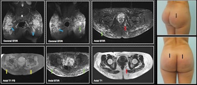 A 28-year-old patient was injected with biopolymers in the gluteal region seven years ago and now is experiencing inflammatory skin changes (blue arrows). MRI shows biopolymers located mainly in the subcutaneous cell tissue of the gluteal region, predominantly in the upper external quadrant in the form of vacuoles (green arrows). There is compromise of the gluteus maximus, where biopolymers are seen adopting a fusiform pattern at this level (blue arrows). There is migration to the left ischioanal fossa (red arrows). The yellow arrows show its hypointense behavior in T1-weighted fat-saturation sequences; the material has a hyperintense behavior in the short-tau inversion recovery sequences.
MRI provides the most information to the radiologist and should be done on all patients, they stated. "It can warn the surgeon about the level of tissue compromise. It shows the affected planes and the approximate amount of biopolymers injected. It demonstrates their areas in the case of material migration."
Additionally, MRI presents the biopolymer in encapsulated form (rounded-oval or elongated vesicles). The round and elongated vesicles can be seen affecting the subcutaneous cellular tissue, so the location is superficial, while the elongated ones are found on deep planes, infiltrating the gluteal fibers.
"Regarding the most frequent areas of migration, we have found that soft tissues of the lumbosacral region are the most frequent, followed by the bilateral ischiorectal region, female external genitalia, and lower limbs," they noted.
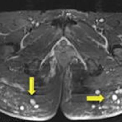
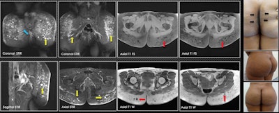 A 35-year-old patient was injected with biopolymers in the gluteal region five years ago. Three years later, changes in the coloration of the skin are visible between the gluteal folds (blue arrow). On MRI, there are multiple vacuoles of hyperintense behavior in the short-tau inversion recovery sequences (yellow arrows) and vacuoles of hypointense behavior in the sequences with T1-weighted information (red arrows), which are compatible with biopolymers located mainly in subcutaneous tissue. In the gluteus maximus muscle, biopolymers are seen in the fusion form (blue arrow). No vacuoles were identified at this level.
A few weeks after placing the biopolymers, liquid forms can be seen that simulate seromas without vesicles. After six to 12 months, seromas and vesicles coexist. After more than 18 months, only vesicles containing the biopolymer material are found. In many patients, there is migration from deep to superficial planes, which, due to their shape and distribution, suggests that lymphatic drainage has occurred.
The length of time that patients carry the biopolymer in the buttocks plays an important role in the way the material will be presented in tissues, the authors wrote.
Other modalities
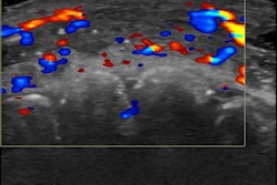
On ultrasound, historically the presence of silicone particles in the subcutaneous tissue has been observed, and it determines heterogeneity and "dirty" acoustic shadow, due to the lower speed of ultrasound conduction.
Although this ultrasound appearance is very characteristic, it is possible any underlying soft-tissue pathology such as tumors, tendon injuries, or bursitis may be hidden. Additionally, ultrasound cannot accurately detect the planes or structures involved and the amounts deposited in a certain area, the researchers explained.
The hypodermis presents a marked diffuse alteration of its ultrasonic structure that compromises it throughout its thickness. Therefore, there is an increase in the echogenicity of the superficial portions and a heterogeneous posterior acoustic shadow that does not allow the visualization of the underlying planes.
"There may be confusion when there is no history of previous exposure, so we must be familiar with the characteristic ultrasound appearance and with the normal appearance of the skin and subcutaneous tissue," Echeverri and colleagues pointed out.
CT is not considered the method of choice in the study of biopolymers because it does not provide adequate anatomical details of the planes involved, and the patient is exposed to radiation. Vacuoles and fusiform forms are usually seen as isodense compared with the muscle, along with partially defined contours, making it difficult to identify in detail their location within the muscle planes.
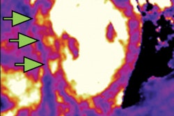
The use of PET/CT is not indicated in the diagnosis for monitoring of biopolymers in the body, but it is good to become familiar with its presentation in this modality, Echeverri and colleagues continued. Biopolymers generate local inflammation, and PET/CT helps to detect those metabolically active tissues, even in the very early stages, and also without clinical manifestations. Although it helps to interpret the anatomy in conjunction with other types of images, it does not give the morphological detail that MRI provides.
X-ray is not a method of choice in the diagnosis or study of biopolymers, but due to the frequency of these esthetic procedures and the widespread use of x-ray, it is good to be familiar with the findings of this modality, the authors concluded.


