Portable MRI can help clinicians quickly and accurately identify intracranial hemorrhages in patients exhibiting signs of stroke -- information that can be potentially lifesaving, according to a study published August 25 in Nature Communications.
Why? Because in patients with stroke symptoms, it can be unclear whether these are caused by a clot that can be treated with blood thinners or by a brain bleed that requires surgery, wrote a team led by Dr. Mercy Mazurek of Yale University in New Haven, CT. Portable, low-field MRI can help quickly clarify the symptoms' cause even in situations where extensive brain imaging isn't available.
"There is no question [portable MRI] can help save lives in resource-limited settings, such as rural hospitals or developing countries," said corresponding author Dr. Kevin Sheth, also of Yale, in a statement released by the university.
Timely brain imaging is critical when patients show signs of stroke, especially because -- if the injury is being caused by intracerebral hemorrhage -- treating the patient with blood thinners is dangerous, the team noted. The American Heart Association recommends that patients suspected of stroke undergo brain imaging on hospital admission before starting treatment with blood thinners.
Noncontrast CT has been the modality of choice for rapid stroke assessment. MRI offers similar benefits for identifying brain hemorrhage, without CT's radiation, but both CT and MRI require transporting patients to the radiology suite, which can use up valuable time in an emergency situation such as stroke, according to the group.
That's where portable MRI shows promise and could improve access to brain imaging and reduce exam time. The system used in the study is a commercially available 0.064-tesla scanner (Hyperfine Research) that weighs 630 kg and can be moved to the patient's bedside.
Mazurek's group compared results of 144 scans acquired with the system at bedside at Yale New Haven Hospital between July 2018 and November 2020 using the portable MRI scanner to results from traditional brain scans (noncontrast CT or 1.5-tesla/3-tesla MRI).
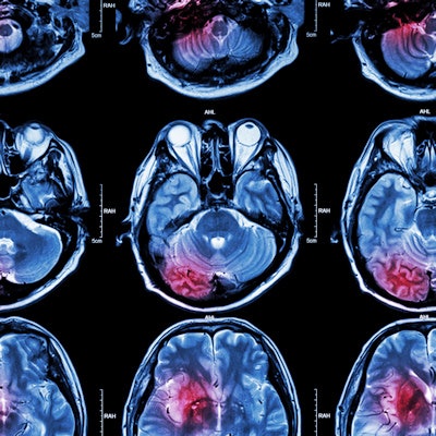
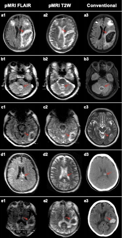 Intracerebral hemorrhage at 0.064-tesla versus conventional imaging modalities (CT or 3-tesla MRI). The first and second columns are low-field FLAIR and T2W images, respectively. The third column is a gold-standard clinical examination for comparison (3-tesla MRI: A3, B3, C3, and E3; CT: D3). (A) Left isointense frontoparietal intracerebral hemorrhage (ICH) with hyperintense rim and bilateral frontal hematomas. (B) Bilateral isointense cerebellar ICH with hyperintense rim. (C) Left hypointense occipital lobe ICH with hyperintense rim. (D) Left homogenous, hyperintense ICH in corpus callosum. (E) Left hypointense temporal ICH with hyperintense rim. Images and caption courtesy of Mazurek, M.H., Cahn, B.A., Yuen, M.M., et al. and Nature Communications through Creative Commons Attribution 4.0 International License.
Two radiologists evaluated the 144 portable MRI scans taken. Of these, 56 were intracerebral hemorrhage, 48 were acute ischemic stroke, and 40 were healthy controls.
The investigators found the following:
- Of the 144 exams, the readers correctly classified 130 as positive or negative for intracerebral hemorrhage, for an overall accuracy rate of 90.3%.
- Of 56 intracerebral hemorrhage cases, the readers identified 45, for a sensitivity rate of 80.4%.
- The readers correctly identified as blood-negative 85 of 88 ischemic stroke and healthy control cases, for a specificity of 96.6%.
- Total imaging time for a portable MRI exam was 30 minutes, compared with 67 minutes for a conventional MRI exam.
"Our results suggest that [portable MRI]-based neuroimaging assessments are a point-of-care solution that could be useful in a broad range of clinical settings for diagnosis and evaluation," Mazurek and colleagues wrote.
Going forward, the team plans to explore whether portable MRI could also help diagnose and track head trauma, brain tumors, and to evaluate brain health in people with conditions such as high blood pressure, the university said.
Disclosure: The study was funded by the American Heart Association, the U.S. National Institutes of Health, and Hyperfine Research.


