MRI shows higher sensitivity than x-ray when it comes to identifying complications from replacement of the patellar component of the knee, according to a study published March 22 in Radiology.
The findings offer clinicians and their patients a more effective way to evaluate the success of total knee replacement, wrote a team led by Dr. Yoshimi Endo of the Hospital for Special Surgery in New York City.
"In addition to the superb depiction of soft tissue including synovial abnormalities at MRI, the ability to evaluate the implant-bone interface and assess loosening are additional compelling reasons why an MRI examination should be considered for all problematic total knee arthroplasty," the group noted.
Loosening of knee replacement components can cause pain, and it may indicate that additional surgery is needed, Endo's group explained. But which imaging modality can best evaluate knee replacement component loosening has remained unclear.
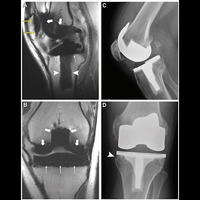
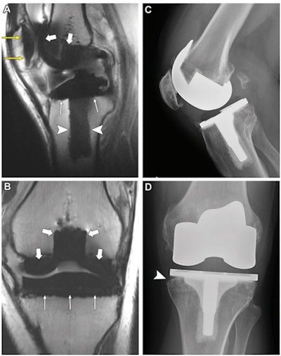
 (A) Sagittal noncontrast-enhanced intermediate-weighted multiacquisition variable resonance imaging combination selective MRI scan (repetition time msec/echo time msec, 4000/7.9) in the midline knee and (B) coronal noncontrast-enhanced intermediate-weighted fast spin-echo MRI scan (4057/32) in the anterior knee in a 51-year-old man show normal interfaces along the patellar component (yellow arrows), femoral component (thick white arrows), and the tibial component, including the tibial tray (thin white arrows) and the keel (arrowheads). (C) Lateral and (D) anteroposterior radiographs in the same knee show normal appearance of the components except for minor bone resorption along the medial tibial tray (arrowhead). Images and caption courtesy of the RSNA.
Endo and colleagues sought to compare the performance of x-ray and MRI for identifying knee replacement component loosening via a study that included 114 patients who had undergone knee replacement surgery (116 knees) between July 2018 and June 2019.
Patients were imaged with both MRI and x-ray, and of the total number of knees, 52.6% had at least one loose component, the team noted. The researchers assessed the type of the interface between the knee replacement component and bone (normal, fibrous membrane, fluid, or osteolysis), the percentage integration of this connection (less than 33%, 33% to 66%, or more than 66%), and the presence of any bone marrow edema; they then compared the sensitivity and specificity of MRI to x-ray for these measures, using surgical findings as reference.
The investigators found that knee replacement component loosening was associated most with the following characteristics:
- Poor (less than 33%) bone integration (odds, 20.4)
- Fluid interface (odds, 20.1)
- Lack of any normal interface (odds, 11.8)
The study also demonstrated that MRI showed higher sensitivity that was statistically significant compared to x-ray for identifying patellar component loosening, although its specificity was lower.
| X-ray vs. MRI for identifying knee replacement patellar component loosening | |||
| X-ray | MRI | p-value | |
| Sensitivity | 31% | 84% | < 0.001 |
| Specificity | 96% | 85% | 0.003 |
Finally, interreader reproducibility between MRI and x-ray for identifying knee replacement component loosening was "substantial to excellent" (0.67 to 0.96), the group found.
The study results highlight the role MRI can play in monitoring the success of total knee replacements, according to the authors.
"Higher sensitivity of MRI for patellar component loosening compared with radiography, together with its superb soft-tissue evaluation, make it an ideal imaging modality for assessing the problematic total knee arthroplasty," they concluded.


