

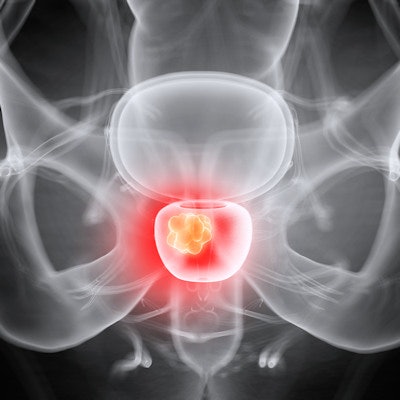
A new microstructure imaging technique, hybrid multidimensional MRI (HM-MRI), could enable better targeting of areas in the prostate that need to be biopsied, according to a presentation at the American Roentgen Ray Society annual meeting in Honolulu.
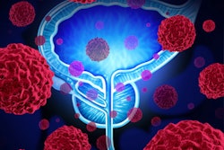
"This study validates [that an] HM-MRI based tool can identify clinically significant prostate cancer -- equal to or more than Gleason 3 + 4 -- more reliably than random biopsy and/or areas detected by a radiologist in patients undergoing MR-ultrasound fusion biopsy," presenter Aritrick Chatterjee, PhD, of the University of Chicago told session attendees.
HM-MRI depicts volumes of tissue components such as the stroma, epithelium, and lumen by "fitting the MRI data into a three-compartment signal model," Chatterjee explained.
He and colleagues conducted a study that included 85 patients with a mean prostate-specific antigen (PSA) score of 8.1 ng/ml; median time between MR imaging and biopsy was seven days. Patients underwent both a standard prostate MRI and an HM-MRI exam; all had a transrectal ultrasound (TRUS)-guided biopsy. The median time between MR imaging and biopsy was seven days.
The group found that HM-MRI had higher sensitivity per tumor of the prostate but not per patient. It had a lower negative predictive value on a per-patient basis.
| Comparison of mpMRI and HM-MRI for identifying clinically significant prostate cancer | ||
| Measure | mpMRI | HM-MRI |
| Per-patient | ||
| Sensitivity | 100% | 90% |
| Specificity | 15% | 40% |
| Positive predictive value | 41% | 47% |
| Negative predictive value | 100% | 88% |
| Accuracy | 46% | 58% |
| Area under ROC curve | 0.58 | 0.65 |
| Per-tumor | ||
| Sensitivity | 70% | 78% |
| Positive predictive value | 22% | 34% |
| By sextant (i.e., from ultrasound-guided biopsies of six different areas of the prostate) | ||
| Sensitivity | 50% | 68% |
| Specificity | 82% | 86% |
| Positive predictive value | 19% | 29% |
| Negative predictive value | 95% | 97% |
| Accuracy | 79% | 85% |
| Area under the ROC curve | 0.66 | 0.77 |
The findings highlight the fact that radiologists need to consider the HM-MRI data with care.
"[We] have to look at these results very carefully and consider all three," he said.
HM-MRI shows promise for improving MR-ultrasound fusion biopsy results by offering more accurate clinical information compared to PI-RADS-based radiologist evaluation, Chatterjee concluded.
"The advantage of this tool is its ability to provide automated, quantitative, reproducible results," he said.
.fFmgij6Hin.png?auto=compress%2Cformat&fit=crop&h=100&q=70&w=100)


.fFmgij6Hin.png?auto=compress%2Cformat&fit=crop&h=167&q=70&w=250)


