Microultrasound may be a suitable alternative to MRI for predicting pathological tumor volume in prostate cancer lesions, according to research published March 13 in Urologic Oncology: Seminars and Original Investigations.
A team led by Arnas Rakauskas, MD, from Lausanne University Hospital in Switzerland found that while microultrasound can predict prostate volume more accurately than MRI, the latter can identify more tumors. However, both modalities tend to slightly underestimate pathological tumor and prostate volume.
“Our study shows that both microultrasound and MRI can be useful in planning focal treatments or radical prostatectomy strategy,” the Rakauskas team wrote. “However, the underestimation of real tumor size in imaging should always be considered when planning treatment margins.”
Researchers continue to explore ways that individualized prostatectomy and focal therapy can lessen treatment-related toxicity while benefitting prostate cancer patients. Multiparametric MRI and microultrasound, the latter using high frequencies of 29 MHz, have emerged as imaging modalities that can stratify risk in these patients.
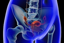
 Images show the measurement of microultrasound prostate and tumor volume using ExactVuTM dedicated software. Images are available for republishing under a Creative Commons license (CC BY 4.0).
Rakauskas and colleagues evaluated the accuracy of microultrasound in predicting the pathological tumor volume of single lesions against MRI in a study that included 65 men with a total of 104 lesions. They also compared the ability of microultrasound to predict prostate volume against MRI.
Microultrasound found a larger median tumor size than T2-weighted MRI, diffusion-weighted imaging (DWI), and apparent diffusion coefficient (ADC) sequences. However, these were all short of the median tumor size detected by pathology.
| Comparison between modalities in predicting prostate tumor volume | |
|---|---|
| Modality | Median tumor size (in ml) |
| Pathology | 1.2 |
| Microultrasound | 1.05 |
| DWI | 0.94 |
| ADC | 0.86 |
| T2-weighted MRI | 0.73 |
On average, microultrasound underestimated pathological tumor volume by 0.15 ml (p < 0.01) while DWI underestimated tumor size by 0.26 ml (p < 0.01). MRI and microultrasound underestimated the pathological prostate volume by 6 ml (p < 0.01) and 3 ml (p = 0.47), respectively.
“The amount of underestimation tended to increase with larger tumors for both imaging modalities with the smallest deviations for microultrasound,” the team wrote.
MRI detected 27% more total tumors than microultrasound. However, if only posterior tumors were counted, microultrasound had a comparable detection rate to the MRI (74% versus 71%, respectively), the researchers reported.
The study authors further highlighted that their results suggest that knowing about imaging underestimation of actual tumor sizes could be useful when planning treatments with any modality.
“Potentially, our results and results from similar studies could be used in generating AI models for live prostate lesion estimation during treatments,” they wrote.
The full study can be accessed here.