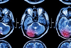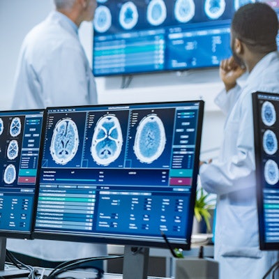
Abbreviated MRI protocols are effective for the diagnosis of emergent conditions, according to a review published May 11 in RadioGraphics.
A team led by Dr. Laura Eisenmenger of the University of Wisconsin in Madison calls these shortened protocols focused abbreviated survey techniques, or FAST, and noted that they can make a difference in patient care across a variety of indications.
"Rapid MRI protocols (including our in-house FAST protocols and more generally all abbreviated MRI protocols) are designed to confidently include or exclude such actionable findings as hydrocephalus, syrinx, seizure, tumor, infection, traumatic injury, and vascular malformations, among others," the group wrote.
MRI has grown in both its technical development and clinical applications, particularly neuroradiology. But its use can be limited by long exam and image interpretation times, the team explained. To address these limitations, researchers have worked to develop shorter MRI protocols that make the modality more effective in urgent care situations that require neuroimaging.
The University of Wisconsin developed three rapid MRI protocols, originally for stroke evaluation but now also used for other applications such as brain and spine, according to the team. The three protocols include the following:
- FAST stroke with contrast. "[This] is our default choice for acute neurologic emergency MRI, including that in stroke workup ... [and] involves comprehensive diffusion imaging and perfusion imaging, which are performed within the first few minutes of image acquisition," the authors wrote.
- FAST spine protocol. "While a more traditional total spine MRI protocol can take up to 90 minutes, our FAST spine protocol can be completed in less than 20 minutes," they explained. "Following localizer image acquisitions, sagittal T1-weighted three-dimensional [fast spin echo] and sagittal two-dimensional short [tau] inversion-recovery (STIR) MR images of the cervical, thoracic, and lumbar regions are obtained."
- "Quick brain" MRI. "[This exam] consists mainly of three two-dimensional [single-shot fast spin echo] acquisitions, which yield sagittal, axial, and coronal whole-brain T2-weighted images within a total table time -- including positioning, pre-imaging, section prescription, and the combined imaging times for all three planes -- of less than four minutes," the group wrote.
The protocols have had a positive effect in the emergency department, according to Eisenmenger and colleagues.
"Given the urgency implicit with stroke and other emergent neurologic conditions and our desire to limit the time spent performing screening examinations, our FAST protocols are targeted to achieve a total imaging time of less than 10 minutes ... and a total room time of less than 15 minutes," they wrote. "FAST protocols have become critical diagnostic screening tools for our emergency department, allowing confident treatment disposition for positive cases and often welcome discharge for negative results."


