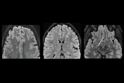
TORONTO - Diffusion spectrum imaging (DSI) is the preferred choice for MR imaging of mild traumatic brain injury, suggest findings presented June 2 at the International Society for Magnetic Resonance in Medicine (ISMRM) annual meeting.
The results were part of a poster session for the International Society of MRI Radiographers & Technologists (ISMRT). Presenter JinRui Zhang, PhD, from Chongqing Emergency Medical Center in China shared research that compared DSI with diffusion tensor imaging (DTI), finding that DSI demonstrates more accurate imaging in this area.
"This modality allows precise diagnosis of mild traumatic brain injury in daily clinical practice, thereby improving patient outcomes," Zhang said.
DSI maps fiber architectures in brain tissues. The researchers highlighted that this method is a valuable tool for exploring brain structures and bridges micro- and macroscopic scales. DTI, meanwhile, detects how water travels along white-matter tracts in the brain.
Zhang and colleagues sought to compare the two modalities, evaluating their respective diagnostic value for assessing microstructural brain tissue changes in patients with mild traumatic brain injury. They included 12 patients in their study; six had acute mild traumatic brain injury and six were healthy volunteers. All study participants underwent brain MRI. The investigators matched the patients for age, gender, and handedness and gathered data from both modalities from brain images after transforming the data format and adjusting for head movement correction.
The team also used an analysis method called tract-based spatial statistics for the study. This method does not need a preset region of interest and helps better align and register the main white-matter fiber tracts of different patients.
Zhang and colleagues found that DTI had a higher mean diffusivity value in the brain injury group than the volunteer group when it came to the posterior limb of the right internal capsule (p < 0.05). However, after implementing tract-based spatial statistics, both DSI and DTI had comparable effectiveness in the posterior limb of the right internal capsule.
However, DSI showed advantages in other areas. The researchers also reported that doctors "subjectively" reported that DSI had higher image signal-to-noise ratio, clearer image quality, small whole-image distortion, and smaller mass area than those of DTI.
The researchers suggested that DSI could make way for further exploration and integration of multi-scale analytical studies at the cellular and subcellular levels.
"In addition to fine display of cross fibers and better guidance of clinical surgery, DSI tracking technology can show the patterns of neural circuits between cerebellar cortex, deep cerebellar and brainstem nuclei, and thalamus, revealing the complex network connection of the cerebellum," they concluded.


