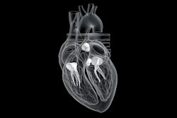
Cardiac MRI shows that higher body mass index, hypertension, and higher physical activity level are associated with higher trabeculated left ventricular (LV) mass, researchers have reported.
And although higher trabeculated left ventricular mass is not "in itself pathologic," it bears tracking, wrote a team led by Nay Aung, MD, of Queen Mary University of London in the U.K. The study findings were published April 2 in Radiology.
"Future studies should evaluate the long-term impact of cardiovascular risk factors on changes in trabecular architecture and subsequent prognostic implications in both individuals with healthy hearts and those with cardiomyopathies," the group noted.
The extent of left ventricular trabeculation and its relationship with cardiovascular risk factors is unclear. Aung's team used data from U.K. Biobank cardiac MRI scans taken between 2006 and 2010 to evaluate any associations between cardiovascular risk factors and trabeculated left ventricular mass. The team applied a deep-learning algorithm to automatically segment trabeculations and compared a cohort of healthy white adults without cardiovascular disease risk factors (7,384 individuals) and a cohort of white adults with preexisting cardiovascular disease risk factors (such as high blood pressure, high cholesterol, diabetes, or smoking; 28,672 individuals).
The researchers found the following:
- Higher body mass index, hypertension, and higher physical activity level were associated with higher trabeculated left ventricular mass.
- In the reference group, the median trabeculated left ventricular mass was 6.3 g for men, and 4.6 g and 4.6 g for women.
"Deep learning-based automated segmentation enabled accurate, high-throughput quantification of left ventricular trabeculation and papillary muscle in a large population-based biomedical database, the U.K. Biobank," the group concluded.
The full article can be found here.


