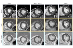
Cardiovascular MRI combined with an AI model can predict heart failure risk in the general population by estimating pressure in the heart, according to research published August 12 in the journal European Society of Cardiology Heart Failure.
A team led by Ross Thomson, of Queen Mary University of London in the U.K., and senior author Pankaj Garg, MD, of the University of East Anglia in Norwich, U.K., conducted a study that found that individuals with higher heart pressure measured by MRI had a fivefold increased risk of developing heart failure over six years.
"One of the most significant findings of this study is that MRI-derived pressure measurements can reliably predict if an individual will develop heart failure," Garg said in a statement released by the University of East Anglia.
The prevalence of heart failure is increasing, in part due to an aging population, the group explained. A typical sign of the condition is raised left ventricular filling pressure, which is estimated from pulmonary capillary wedge pressure (PCWP), usually via cardiac catheterization. But cardiovascular MRI is becoming an increasingly valuable tool for assessing patients with suspected heart failure, in part because it is less invasive.
Yet although research has shown that cardiac MRI can estimate pressure in the heart -- which is linked to symptoms of heart failure -- it has been unclear whether this information can predict heart failure risk in a general population, Thomson and colleagues explained.
To address the question, the team conducted a study that included data from more than 39,000 U.K. Biobank participants, using AI techniques with MRI to estimate pressure in the heart. (U.K. Biobank is a biomedical database that consists of deidentified genetic, lifestyle, and health information and biological samples from half a million U.K. participants.) The authors then evaluated each individual's risk factors (i.e., being male, over 70 years of age, having high blood pressure, obesity, and alcohol consumption) for increased pressure in the heart and the likelihood of developing heart failure over a six-year follow-up period.
Thomson and colleagues used AI to calculate cardiovascular magnetic resonance-modeled PCWP, incorporating data regarding left atrial volume, left ventricular mass, and sex. They investigated any links between cardiovascular risk factors and high cardiovascular MRI-modeled PCWP (that is, more than or equal to 15 millimeters of mercury [mmHg]) and assessed the influence of these risk factors and high heart pressures on incident heart failure and major adverse cardiovascular events (MACE). (The researchers defined incident heart failure symptoms and signs of the condition that required patients to be admitted to the hospital, and defined MACE as the composite outcome of nonfatal myocardial infarction, nonfatal stroke, and cardiovascular death.)
The group found that clinical characteristics associated with raised cardiovascular MRI-modeled PCWP included the following:
- Hypertension (odds ratio [OR] 1.57, with 1 as reference)
- Body mass index (OR, 1.57)
- Male sex (OR, 1.37)
- Age (OR, 1.33)
- Regular alcohol consumption (OR, 1.1)
The team also found that cardiovascular MRI-modeled PCWP was independently associated with incident heart failure (hazard ratio, 2.91), and MACE (hazard ratio, 1.48).
The study results could help clinicians better track patients' heart health.
"Cardiovascular MRI-modeled PCWP should be incorporated into routine cardiovascular MRI reports to guide heart failure diagnosis and further management," the authors concluded.
The complete study can be found here.


