A Swiss research team has developed and tested an AI model that automatically segments anatomic structures on MR images independent of sequence, according to a study published February 18 in Radiology.
The study results suggest that the model, called TotalSegmentator MRI, could improve radiology department workload, said senior author Jakob Wasserthal, PhD, of University Hospital Basel in Switzerland.
"MRI images have traditionally been manually segmented, which is a time-consuming process that requires intensive effort by radiologists and is subject to interreader variability," he said. "Automated systems can potentially reduce radiologist's workload, minimize human errors and provide more consistent and reproducible results."
An automated segmentation tool called TotalSegmentator CT was released in August 2023, and since then there has been demand for a similar tool that can be applied across all MRI sequences and anatomic structures, the group explained. To this end, D'Antonoli and colleagues built TotalSegmentator MRI, which they based on nnU-Net. They trained the model to produce sequence-independent segmentations of major anatomic structures using a dataset of 616 MR and 527 CT exams (total scans, 1,143). Of these, 1,088 (both CT and MRI exams) were used in a training set that included segmentations of 80 anatomic structures commonly used for measuring volume, characterizing disease, surgical planning, and opportunistic screening, while 55 (MRI exams only) were included in an internal test set. The team then assessed the model's performance using Dice scoring.
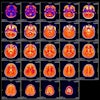
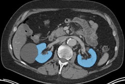
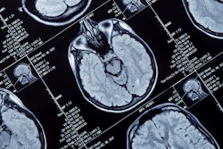
 Example MRI scans in the training dataset. Since images were randomly sampled from clinical routine, the dataset contains a wide variety of different contrasts, pathologies, and image types. Images and caption courtesy of the RSNA.
The authors reported that TotalSegmentator MRI had a Dice score of 0.839 for the 80 anatomic structures in the internal test set and that it outperformed two other models (with a Dice score of 0.862 vs. 0.759 for 40 anatomic structures and a Dice score of 0.838 vs. 0.56 for 13 anatomic structures).
In addition to research and AI product development, the model could potentially be used for treatment planning, monitoring disease progression, and opportunistic screening, Wasserthal said.
"To our knowledge, our model is the only one that can automatically segment the highest number of structures on MRIs of any sequence," he said. "It's a tool that helps improve radiologists' work, makes measurements more precise, and enables other measurements to be done that would have taken too much time to do manually."
Tools like TotalSegmentator MRI show promise for streamlining radiology department operations, wrote Felipe Kitamura, MD, PhD, of the Universidade Federal de São Paulo in Brazil, in an accompanying commentary -- although challenges to incorporating AI in this way remain.
"The appeal of such models lies in their potential to reduce the high costs and logistic complexities associated with collecting, annotating, and curating medical data for specialized segmentation tasks," Kitamura noted. "Rather than painstakingly compiling thousands of annotated studies for each new organ or pathology, a single foundation model could be adapted with relatively few new examples. However, realizing this vision depends on resolving challenges such as data governance, patient privacy, and robust data labeling."
TotalSegmentator MRI can be accessed here.
The complete study can be found here.