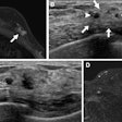
While ultrasound of the breast is helping to determine malignancy of lesions seen on mammography, it still doesn't preclude large numbers of women from undergoing unnecessary biopsy of masses that ultimately prove to be benign.
That's why a number of researchers are looking at various ultrasound enhancements that may spare more women from unneeded trauma. Enhancements including tissue harmonic imaging, ultrasound contrast, and computer-aided evaluation of sonographic breast images, are all being examined as ways of improving the modality's predictive capability. The technologies were scrutinized at a scientific session at last month's Radiological Society of North America meeting.
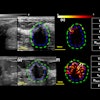
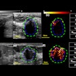
Even older technologies such as color Doppler ultrasound may have untapped potential to differentiate likely benign from malignant lesions, according to Dr. Hye-Young Choi of the department of diagnostic radiology at Ewha Women's University in Seoul, South Korea.
In a retrospective analysis, Choi looked at 129 consecutive cases of suspicious lesion to determine the location, pattern and shape of the Doppler signals that were observed. Her analysis found that lesions on the periphery of the breast were more likely to be benign, while branching tumors which were highlighted by the color Doppler were more likely to be malignant.
Choi also concluded that power Doppler was more useful than color Doppler for detection of vascular signals.
Other technologies
Radiologists in one small study said they preferred tissue harmonic imaging (THI), a proprietary ultrasound enhancement, because it appeared to improve margin analysis, conspicuity, and overall quality of breast images compared to standard ultrasound. Dr. Eric Rosen, a researcher at Duke University Medical Center in Durham, NC, said the narrower beam used in THI may result in better sonographic pictures.
Rosen's study looked only at radiologists' preferences for THI or standard sonographic images, and did not examine whether THI resulted in more accurate determination of solid or cystic lesions. However, he still concluded that THI may improve ultrasound's sensitivity and specificity in evaluating breast lesions.
Researchers in Germany have also looked at use of the ultrasound contrast agent Levovist to help distinguish benign from malignant breast lesions. Dr. Jens Uwe Blohmer, from the Frauenklinik Charite, Berlin, administered Levovist intravenously to 22 patients with mammary carcinoma and to 11 women with benign breast lesions.
By looking at the intensity of the sonographic signal, Blohmer said doctors might be able to determine if the lesion is malignant. In his experiments, the greater the time to peak brightness, the more likely the tumor was benign. The average time to peak brightness in benign lesions was 42 seconds, compared to 26 seconds for malignant tumors. Blohmer suggested the use of the agent "might be useful in differential diagnosis."
And then there are the efforts to train computers to read and analyze breast images with greater accuracy than can typically be achieved by human radiologists.
Dr. Joseph Lo, PhD, an assistant professor of radiology also at Duke University, input 192 sonograms into an artificial neural network (ANN) and reported that the computer program was able to identify all 71 malignancies on the breast images.
The ANN was also able to exclude from biopsy 48 of 121 benign tumors, achieving a total positive predictive value (PPV) of 55%, better than the 48% PPV achieved by radiologists who originally read the images.
In one case, a radiologist who looked at an ultrasound image rated the lesion as highly likely to be malignant. The ANN disagreed, reporting a less than 20% chance that the tumor was malignant. The result: It was benign.
Lo noted that the computer made its calls based solely on seven morphological characteristics seen on ultrasound images (mass shape and margin, lesion echogenicity and echotexture, acoustic transmission, and presence of an echogenic pseudocapsule or visible calcification within a lesion). In contrast, the radiologists had additional input for their determinations, including previous sonograms, mammograms and patient history.
Lo plans to continue adding cases to the ANN, which has the capability of learning as it gathers more information.
But while these studies showed greater potential for ultrasound, none offered conclusive evidence that their respective technological enhancements could be applied to clinical practice without a lot more investigation.
"These are really works in progress," said Dr. Ellen Mendelson, chief of women's imaging and director of the Breast Center at Western Pennsylvania Hospital, Pittsburgh. She was the moderator of the RSNA session that discussed the various studies.
"What I saw here was an excellent manifestation of increasing interest in ultrasound, and what it might and might not be able to do," Mendelson said.
By Edward Susman
AuntMinnie.com contributing writer
January 5, 2000
Copyright © 2000 AuntMinnie.com


