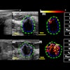
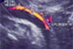
(Radiology Review) Giant cell tumors of the flexor tendons of the fingers appear as homogeneous masses with reduced echogenicity (hypoechoic) on ultrasound imaging, according to research recently published in American Journal of Roentgenology (AJR 2004, Vol. 183, pp. 337-339). Also known as localized nodular tenosynovitis, giant cell tumors of the tendon sheath rarely become malignant, Dr. William D. Middleton and colleagues said.
Radiologists at Washington University School of Medicine, St. Louis, performed a retrospective search of ultrasound studies of the hand and wrist published over a five-year period until December 2001. They found 429 such studies, with 12 cases of confirmed giant cell tumor of the hand.
Sonographic features of interest included tumor site, tumor size, echogenicity, bone erosions, calcification, attenuation characteristics, and presence of cysts. The degree of contact with the tendon and amount of lesion blood flow also were assessed.
Of the 12 patients studied, "ten patients presented with a painless mass, two with a painful mass," they reported. "Tumor size ranged from 3 to 15 mm with a mean of 9.7 mm."
Review showed that all tumors were in contact with a tendon and all showed reduced echogenicity. Ten were homogeneous and two were heterogeneous. Increased through transmission was demonstrated in four cases, but the remainder showed normal attenuation characteristics. Bone erosions at the metacarpal-phalangeal joint were shown in two cases. Ten of the lesions involved the flexor tendon (volar surface) and two involved the extensor tendon (dorsal surface).
"In all cases, it was noted that the tumors did not move with the tendon when the affected digit was flexed or extended," they said. Color and pulsed Doppler demonstrated internal vascularity in 11 tumors and five lesions showed increased vascularity compared with the surrounding tissue.
The importance of differentiating giant cell tumors and ganglionic cysts was noted. Treatment for both was surgical resection; however, ganglionic cysts also required steroid injection, the authors said. "Giant cell tumors of tendon sheath have a characteristic sonographic appearance and occur in predictable locations." They appear as solid, hypoechoic lesions adjacent to the flexor tendons that show blood flow on color Doppler, they said.
Giant Cell Tumors of the Tendon Sheath: Analysis of Sonographic Findings
William D. Middleton, et al
The Mallinckrodt Institute of Radiology
Washington University School of Medicine
510 Kingshighway, St. Louis, MO 63110, USA
AJR 2004 (August); 183:337-339
By Radiology Review
August 9, 2004
Copyright © 2004 AuntMinnie.com


