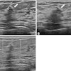
BOSTON - Ultrasound elastography is superior to conventional ultrasound in characterizing breast lesions and could help reduce unnecessary breast biopsies, according to a study presented by researchers from Singapore on Wednesday at the American Roentgen Ray Society (ARRS) meeting.
"Elastographic ratios perform better than sonographic features in distinguishing malignant from benign beast lesions," said presenter Dr. Llewellyn Sim from Singapore General Hospital.
Although ultrasound is commonly used as an adjunct to x-ray mammography for diagnosing breast cancer, the modality suffers from low specificity. To see if elastography could improve upon conventional ultrasound's performance, the Singapore researchers sought to compare the technique with ultrasound features in assessing breast lesions.
The study team evaluated 99 women with 110 sonographically visible breast lesions from September 2007 to March 2008. Patients were evaluated independently by three breast radiologists with conventional ultrasound, elastography, and a combination of the two modalities.
Both B-mode and elasticity images were acquired using an Antares ultrasound scanner (Siemens Healthcare, Erlangen, Germany). BI-RADS scores were assigned to standardize ultrasound interpretation, while elastograms were classified as benign, malignant, or equivocal based on the strain pattern and the length and area ratios, Sim said. The results were then correlated with histopathology to evaluate performance.
Of the 100 breast lesions, 26 were malignant and 84 were benign. Elastography and the combination of ultrasound and elastography both yielded significantly better results than ultrasound alone (p < 0.0005), Sim said.
|
From receiver operator characteristics analysis, the researchers determined that the optimal length and area ratios for distinguishing benign and malignant lesions on elastography were both 1.1, Sim said.
In other findings, the elastography area and distance ratios were superior to every sonographic feature for characterizing lesions, Sim said. The area ratios achieved higher sensitivity, specificity, and accuracy than the distance ratio, but the differences were not statistically significant.
In conclusion, breast elastography has a higher sensitivity, specificity, and accuracy than conventional ultrasound, Sim said.
"The use of breast elastography alone or combined with ultrasound provides a more accurate diagnosis of breast cancer," he said. "In terms of clinical significance, the better performance of [breast elastography] improves sonographic diagnosis of breast cancer, reinforces a benign ultrasound finding, and potentially reduces unnecessary biopsies."
A combined algorithm consisting of both elastographic parameters and ultrasound features could increase diagnostic accuracy and also be used in a computer-aided detection program for analysis of breast lesions, Sim said.
By Erik L. Ridley
AuntMinnie.com staff writer
April 29, 2009
Related Reading
Ultrasound elastography shows potential in thyroid nodules, February 11, 2009
Ultrasound elastography shows strength for diagnosing rotator cuff tears, January 15, 2009
Adding sonography to yearly mammograms helps find metachronous cancers, February 25, 2008
Breast sonoelastography aids in evaluating breast lesions, January 28, 2008
Elasticity imaging finds breast tumors based on shape, volume, December 14, 2007
Copyright © 2009 AuntMinnie.com


