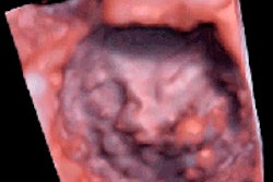
3D transesophageal echocardiography (TEE) is superior to 2D TEE for quantifying the severity of mitral regurgitation, according to researchers from Leiden University Medical Center in Leiden, the Netherlands.
In a paper published online in Circulation: Cardiovascular Imaging, a study team led by Miriam Shanks, MD, concluded that 3D TEE was both feasible and accurate compared to MRI quantitative assessment. The group also found that 3D TEE was less prone than 2D TEE to underestimate regurgitant volume.
The American Society of Echocardiography (ASE) emphasizes that quantitative techniques be used to assess mitral regurgitation to provide risk stratification and clinical management of patients with this condition. However, the standard approach of 2D color-flow and Doppler echocardiography with the proximal isovelocity surface area (PISA) method has been problematic for grading regurgitant volume (marker of volume overload) and effective regurgitant orifice area (descriptor of lesion severity), according to the researchers.
Hoping to make use of recent 3D TEE technology advances, the study team sought to compare the performance of 2D and 3D TEE using MRI as a reference method (Circulation: Cardiovascular Imaging, September 1, 2010).
The researchers studied 30 patients (20 men, 10 women; mean age, 63.3 years) who were clinically referred for TEE and cardiac MRI for assessment of mitral regurgitation. Exclusion criteria included patients with irregular heart rhythm, a history of mitral valve replacement, significant aortic or tricuspid valve regurgitation, or absolute contraindications to TEE and MRI.
All patients received 2D and 3D TEE as well as a cardiac MRI on the same day.
Both 2D and 3D TEE were performed using an iE33 ultrasound scanner (Philips Healthcare, Andover, MA) with an X7-2t fully sampled matrix-array TEE transducer.
After complete 2D, color, pulsed-, and continuous-wave Doppler images were obtained for assessing cardiac structures and function, the researchers quantitatively determined mitral regurgitation severity from the four-chamber views obtained at midesophageal level with zero-degree tilt.
In both TEE methods, mitral effective regurgitant orifice area and regurgitant volume were estimated. Mitral effective regurgitant orifice area was calculated from 3D TEE using planimetry of the color Doppler flow from "en face" views, while regurgitant volume was determined by multiplying the mitral effective regurgitant orifice area by the velocity time integral of the regurgitant jet on the continuous-wave Doppler, according to the authors.
2D measurements of effective regurgitant orifice area and regurgitant volume were generated using the standard proximal isovelocity surface area method.
MR images were captured on a Gyroscan ACS-NT/Intera 1.5-tesla scanner (Philips), with a five-element cardiac synergy coil. Mitral regurgitant volume was determined by subtracting the aortic flow volume from left ventricular stroke volume, according to the researchers.
The group discovered that 2D TEE underestimated the mitral effective regurgitant orifice area by a mean of 0.13 cm2 compared to 3D TEE. 2D TEE was also found to underestimate the regurgitant volume by 21.6% compared to 3D TEE and by 21.3% compared to MRI.
"In contrast, 3D TEE underestimated the [regurgitant volume] by only 1.2% when compared to MRI," the authors noted.
In other findings, the researchers noted that one-third of the patients in grade 1 (moderate regurgitation) and 50% or more of patients in grades 2 and 3 (moderate to severe regurgitation) would have been upgraded to a more severe grade based on the 3D TEE and MRI measurements, compared with what they would have received under 2D TEE.
The study team also detected good intraobserver variability (mean difference of 0.011 ± 0.16 cm2; intraclass correlation coefficient = 0.98) and interobserver variability (mean difference of 0.013 ± 0.14 cm2; intraclass correlation coefficient = 0.98) in a comparison of 3D mitral effective regurgitant orifice area measurements.
"The present study demonstrated that 3D TEE is feasible, and is more accurate in the quantitative assessment of both functional and organic mitral regurgitation when compared to 2D TEE," the authors concluded. "Given the potential strengths of 3D echocardiography, prospective studies to determine the importance of the 3D data for the clinical outcome may be needed."
By Erik L. Ridley
AuntMinnie.com staff writer
September 10, 2010
Related Reading
Transesophageal echo informative in cryptogenic stroke, April 23, 2009
Real-time 3D TEE helps guide heart-repair procedures, September 4, 2008
Echocardiography predicts pulmonary artery pressure in ventilated patients, March 25, 2008
3D echo effective in aortic stenosis appraisal, July 3, 2007
Transesophageal echo spots pulmonary vein stenosis, June 4, 2007
Copyright © 2010 AuntMinnie.com


