Thursday, December 1 | 11:10 a.m.-11:20 a.m. | SSQ03-05 | Room E353A
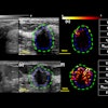
An Italian team believes that real-time shear-wave elastography can improve upon the limitations of transient elastography in detecting liver fibrosis.Transient elastography has emerged as the noninvasive reference test for the liver and is entering clinical practice in Europe. While the technique has been successful in detecting severe fibrosis, transient elastography has struggled in detecting significant fibrosis, a critical factor to starting treatment, said presenter Dr. Giovanna Ferraioli of the University of Pavia.
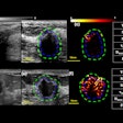
As a result, the researchers wanted to determine if shear-wave elastography could improve upon some of transient elastography's limitations. In a study comparing shear-wave elastography performed on an Aixplorer system (SuperSonic Imagine) and transient elastography performed using a FibroScan system (Echosens), the diagnostic performance of shear-wave elastography compared favorably with transient elastography, according to the researchers.
Shear-wave elastography appears to be an accurate technique for detecting and staging liver fibrosis, according to the results, Ferraioli said.
"[Shear-wave elastography] seems a promising noninvasive imaging technique in the management of chronic liver diseases that could be used to evaluate when to start antiviral or antifibrotic therapies," Ferraioli said. "Moreover, because of its noninvasiveness, [shear-wave elastography] can be repeated over time, thus allowing disease and treatment monitoring."


