Friday, December 2 | 11:20 a.m.-11:30 a.m. | SST08-06 | Room E351
In this Friday session, a U.K. team will show how real-time tissue elastography can increase operator confidence and influence patient management for evaluating intratesticular lesions.Clinical assessment of the testes relies heavily on palpation to identify and evaluate pathological processes. However, the advent of elastography has enabled the determination of tissue stiffness with imaging: The researchers wanted to investigate whether the technique could be used in testicular imaging and whether it could be employed to sonographically "palpate" a lesion within the testis, said presenter Dr. Aarti Shah of King's College Hospital.
Because ultrasound is the main tool for imaging the testes, any technique that can be performed as part of a routine exam to improve sensitivity and specificity offers tremendous potential, she said.
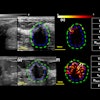
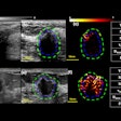
In a two-year study, 28 patients with focal intratesticular lesions received real-time tissue elastography on an HI Vision 900 ultrasound scanner (Hitachi Medical) with a 14-6 MHz linear-array transducer. The method yielded 83% sensitivity for detecting malignant lesions.
The researchers concluded that elastography can be used to assess small, indeterminate, intratesticular lesions to characterize their hardness or softness using a color scale.
"When used in conjunction with B-mode ultrasound, elastography can increase operator confidence and perhaps avoid unnecessary biopsy or even orchiectomy," Shah said.


