Tuesday, December 2 | 11:10 a.m.-11:20 a.m. | VSMK31-11 | Room E450A
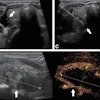
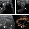
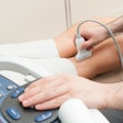
Supersonic shear imaging has the potential to characterize changes in the Achilles tendon brought on by aging and disease, and it could perhaps decrease the risk of injury, according to this presentation to be given on Tuesday.Researchers from the University of Wisconsin School of Medicine and Public Health used the technology to evaluate age-related changes of the Achilles tendon and calf muscle strain in adults. The study consisted of 10 young and 10 middle-aged adults (average age, 27 and 49, respectively).
Lead researcher Dr. Kenneth Lee and colleagues collected shear-wave images from regions of the Achilles tendon, including the muscle-free tendon, the soleus aponeurosis, and the medial gastrocnemius aponeurosis, measuring the shear-wave speeds of each site at three angles: resting, dorsiflexed, and plantarflexed.
The performance of supersonic shear imaging varied depending on imaging location and the ankle position; the technology was most effective when imaging the muscle-free tendon and increasing ankle dorsiflexion, the group found.
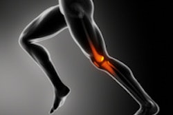
The results suggest that the elasticity of the Achilles tendon decreases with age. However, with middle-aged adults, elastography showed greater flexibility near the muscle-tendon junction, which could increase the risk of injury such as commonly seen with "tennis leg," the researchers concluded.

