Sunday, November 29 | 12:30 p.m.-1:00 p.m. | GU200-SD-SUA1 | Lakeside Learning Center, Station 1
Women in poor communities often lack access to basic obstetric imaging. But researchers from the University of Vermont found that even minimally trained sonographers can produce diagnostic-quality obstetric ultrasound images.The study findings suggest that basic ultrasound could improve perinatal outcomes in poor areas, according to poster presenter Matthew LeComte, PhD, and colleagues.
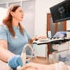
LeComte's group invited women who had an obstetric ultrasound performed by an expert to undergo another ultrasound performed by a less experienced sonographer who conducted anatomically guided sweeps with the ultrasound probe.
These images were then evaluated by two obstetricians and one radiologist and compared with the initial ultrasound. The three readers assessed the visibility of maternal and fetal anatomy, gestational and placental features, and various fetal growth measures, and they ranked their confidence level for each (confident, probable, or uncertain).
The readers evaluated 61 studies, and they described 62% of the sweep images of the fetus as well-visualized. They rated 97% of the reports as confident for confirming pregnancy and 98% as confident for confirming fetal and placental position. Half of the exams were characterized as confident for imaging growth measures of the fetus' cardiac, urinary, abdominal, and neuroanatomy.
The researchers plan to further analyze the study data, but they concluded that even an individual minimally trained as a sonographer -- using only anatomic landmarks -- can generate diagnostic-quality obstetric ultrasound images upon which clinical decisions can be made.

