Tuesday, November 27 | 11:20 a.m.-11:30 a.m. | SSG01-06 | Room S406A
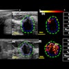
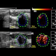
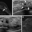
Using a simple five-point technical quality scale can help radiologists better use shear-wave elastography (SWE) data, according to scientific research to be highlighted on Tuesday morning.Presenter Dr. Masoud Baikpour of Massachusetts General Hospital and colleagues conducted a study that included 110 women with 122 breast lesions who underwent SWE and ultrasound-guided biopsy between October 2017 and January 2018. The researchers recorded the maximum, mean, and standard deviation elasticity measurements for each lesion. They also set five SWE technical quality measures:
- Lesion visibility on B-mode images
- Stiffness pattern in the near field of the field-of-view
- Size and location of the field-of-view box relative to lesion
- Heterogeneity, vertical streaks, and absence of color in tissue surrounding the lesion
- Size and location of the region-of-interest circle on the lesion for elasticity measurements
Three readers assessed each SWE parameter on each lesion as having as either low quality (score of 0) or high quality (score of 1). SWE images with total scores of less than 3.3 were classified as low quality, and those above 3.3 were classified as high quality. The team then compared area under the receiver operating characteristic curve (AUC) of SWE for low- versus high-quality images.
Of the 122 lesions, 52% were benign and 48% were malignant. Baikpour's team found that AUC improved in the high-quality image group compared with the low-quality group for the mean elasticity measurement (0.85 versus 0.63, p = 0.009) and the standard deviation elasticity measurement (0.81 versus 0.62, p = 0.040). The AUC for maximum elasticity measurements also increased in the high-quality image group compared with the low-quality image group (0.86 versus 0.71), but it did not reach statistical significance.
"This data will help radiologists to decide in real-time whether or not to trust the information provided by the SWE images -- that is, whether it's OK to take the SWE measurements and color map into consideration if the image is high-quality, or whether it's better to disregard the SWE image if it is low-quality," co-researcher Dr. Shirley Chou told AuntMinnie.com.


