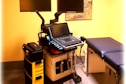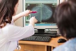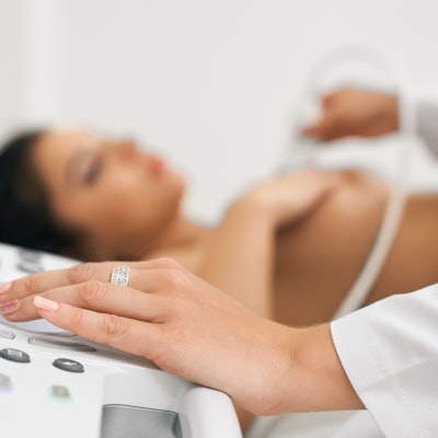
With a national reporting standard for breast density across the U.S. on the near horizon, ultrasound is poised to play a greater role as an adjunct to mammography in screening, along with other imaging modalities.
The U.S. Food and Drug Administration (FDA) proposed a uniform standard for informing patients about breast density after mammography in March 2019, spurred by a new federal law. An update by the FDA standardizing density reporting through the Mammography Quality Standards Act (MQSA) feels overdue and is eagerly awaited by patient advocates.
Breast density is associated with increased risk for cancer and makes it harder to catch cancers on screening mammograms. Dense tissue is made of glands and fibrous tissue and appears white on a mammogram, but cancer is also white, so it is difficult to spot against this backdrop. As a result, many cancers are missed, and women may not get the benefit of early diagnosis and more conservative treatment options. Adding another imaging modality to mammography could help detect cancers in women with dense breasts, but supplemental screening has not been standardized.
Since women are often aware of the limitations of mammography, there has been a push by advocates on the state and national levels for dissemination of information about the effect density has on mammography. According to the advocacy group DenseBreast-info.org, 38 U.S. states and the District of Columbia have passed laws requiring breast cancer facilities to inform women and referring providers about breast density. But the level of information differs significantly; for example, a patient's individual density results may or may not be communicated.
Where ultrasound fits in
Broadly speaking, what type of imaging modality should be used as an adjunct to mammography is subject to debate. In accordance with American College of Radiology (ACR) recommendations, there is general agreement that women who are at high risk of breast cancer should be getting supplemental screening with contrast-enhanced breast MRI, regarded as the most sensitive imaging modality.
High-risk groups include those genetically predisposed for at least a 20% risk of breast cancer. The ACR suggests ultrasound "for those who qualify for but cannot undergo MRI." Reasons for not undergoing MRI include claustrophobia, unwillingness to take contrast, and exceeding table weight limits.
Screening breast ultrasound of the whole breast is commonly used as a supplemental screening tool for evaluating dense breasts after a screening mammogram. Ultrasound is relatively cheap, easy to perform, and does not involve ionizing radiation or injection of intravenous contrast.
The literature suggests that beyond mammography alone, ultrasound identifies an additional 2-3 cancers per 1,000 women screened beyond mammography. Furthermore, more than 85% of cancers seen only on ultrasound are invasive, early-stage, and node-negative.
In the Japan Anti-Cancer Randomized Trial (J-START) study of women with breasts that were heterogeneously dense (BI-RADS category C) or extremely dense (BI-RADS category D), the cancer detection rate was 5 per 1,000 for mammography with ultrasound versus 3.3 per 1,000 for mammography alone. Importantly, the interval cancer rate, which is viewed as a surrogate for mortality, was lower when ultrasound was combined with mammography at 0.5% for the combination compared with 1% for mammography alone.
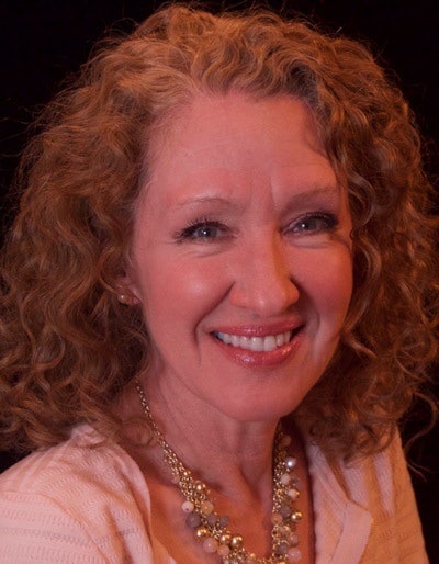 Dr. Liane Philpotts.
"From a screening standpoint, ultrasound makes a lot of sense," commented Dr. Liane Philpotts, breast imaging section chief of the eastern region at Yale School of Medicine.
Yale adopted screening ultrasound as a supplemental screening tool after a state law in Connecticut was passed in 2009 requiring notification of breast density. At Yale-New Haven Hospital where Philpotts practices, mammography technologists were cross-trained in handheld ultrasound in a peer-to-peer training program -- they are finding cancers and ultrasound has proven to be a very useful test in women whose only risk factor is breast density. About three-quarters of patients with dense breasts now get ultrasound as part of their breast screening.
"It's one of those things where once you start doing it, there's no going back," Philpotts said.
 Dr. Paula Gordon.
Ultrasound is very synergistic with mammography: If the two are performed during the same visit, suspicious findings can be quickly clarified by comparing the two studies, she said. At some centers, ultrasound-guided needle biopsy is available on the same day, said Dr. Paula Gordon, clinical professor at the University of British Columbia.
"Being able to do additional tests -- including needle biopsy -- on the same day has huge value," Gordon said.
Some breast imaging experts see a much wider role for ultrasound -- not just a second choice for high-risk women if breast MRI is not available but also as an option for average-risk women who have dense breasts. Gordon said that she believes all women with heterogeneously dense or extremely dense breasts should be offered supplemental screening with ultrasound if they don't qualify for MRI based on their other risk factors.
"Anything less than that is rationing -- and dangerous rationing," Gordon said.
In a University of British Columbia study of women with heterogeneous or extremely dense breasts undergoing supplemental ultrasound screening, 40% of the detected cancers detected were in women whose breasts were heterogeneously dense or extremely dense and who had no family history of breast cancer or other factors for increased risk, Gordon explained. Furthermore, of the cancers detected, 80% were category C. So, limiting ultrasound screening to women with a family history or to only category D would miss most of the cancers, she pointed out.
Limitations of ultrasound
However, there are limitations. Handheld ultrasound is typically performed by technologists and performance is operator-dependent. Standardization nationwide is lacking.
False positives, with associated recalls and biopsies, are also a big concern with supplemental ultrasound screening. In the J-START study, the false-positive rate rose from 8.8% to 12.6% with ultrasound.
A recently published Korean study had a lukewarm assessment of screening ultrasound as an add-on after negative results with digital mammography and digital breast tomosynthesis (DBT) in women with dense and nondense breasts (Radiology 2021;298:568-575). An additional 3.2 cancers were detected per 1,000 screening exams, which the authors described as a "modest gain," while the abnormal interpretation rate, that is the rate of added recalls, was very high at 18%.
 Dr. Habib Rahbar.
In an accompanying editorial, Dr. Habib Rahbar, breast imaging clinical director at the Seattle Cancer Care Alliance, suggested it may be time to "close the chapter" on ultrasound screening trials and focus instead on imaging modalities that have proven to be superior to ultrasound for supplemental screening in women regardless of risk: contrast-enhanced breast MRI and contrast-enhanced mammography (Radiology 2021 Vol. 298 No 3:576-577).
The study and editorial got some pushback in a letter to the editor by Gordon and the University of Pittsburgh breast imaging expert Dr. Wendie Berg, PhD. The starting screening age in the study was young at 30, for reasons that were unclear, and included women with dense and nondense breasts, whereas in the U.S., this is not the population recommended for supplemental screening with ultrasound. All of the additional cancers detected in the study were found in dense breasts and the abnormal call rate may have been inflated by the inclusion of women with nondense breasts, Gordon commented to AuntMinnie.com.
Gordon also questioned the conclusion that the number of additional cancers detected (3.2 per 1,000) only on ultrasound was "modest."
"That's a big deal," Gordon said. "That's life-saving."
Fast MRI is compelling
In an interview, Rahbar said that there is no question that ultrasound is firmly established as a powerful tool for screening, in addition to its role investigating clinical symptoms. However, Rahbar questions investing more and more in research in a tool that seems to max out at about two or three additional cancers per 1,000 women screened and also causes a lot of false positives.
Ultrasound is a great when it comes to morphological detail, but it lacks the functional ability to detect angiogenesis, whereas contrast-enhanced imaging does provide information about blood vessels, improving sensitivity for cancer detection, explained Rahbar, who is also an associate professor of radiology at the University of Washington.
While MRI is very sensitive, it also requires use of intravenously administered gadolinium contrast, and traditionally a long acquisition time of 30-60 minutes, not to mention high cost at over $1,000. But abbreviated MRI acquisition techniques pioneered by Dr. Christiana Kuhl and colleagues in Germany have demonstrated excellent performance on par with full MRI and promise to be a lot less expensive.
In one seminal screening study of women at mild-to-moderate risk after negative mammography and ultrasound, abbreviated MRI found an additional 18.2 cancers per 1,000 screened. This was achieved with an acquisition time of 3 minutes and a reading time of less than 3 seconds (J Clin Oncol 2014;Vol. 32, No. 22: 2304-2310.)
In a prospective, multicenter study of screening in average-risk women with dense breasts, the invasive cancer detection rate was 11.8 per 1,000 women for abbreviated breast MRI compared with 4.8 per 1000 for DBT (JAMA 2020 Vol. 323, No. 8:746-756). For detection of invasive cancer and ductal cancer in situ (DCIS), sensitivity of abbreviated MRI was 95.7% for MRI compared with 39.1% for DBT; specificity was 86.7% versus 97.4%.
Based on the data, the concept of using MRI in non-high-risk patients has "really taken off" and there's a question of whether it could even replace conventional mammography, Rahbar said.
Another emerging screening test is contrast-enhanced mammography (CEM), which uses iodinated contrast. CEM is a fast test with similar efficacy but lower expense than MRI. Like ultrasound, MRI and CEM come with false positives, but the yield of additional cancers detected is a lot higher.
Rahbar believes that the most appropriate use for ultrasound is in women at high risk who cannot tolerate MRI. It's also a good modality for those who have dense breasts and who don't fall into other risk categories who would like supplemental screening and don't want MRI, he said. Where Rahbar practices, screening ultrasound is not generally provided unless these criteria are met.
Some breast imagers who had mixed views on ultrasound for supplemental screening have been gravitating to MRI due to the high sensitivity and promising early study results, commented Dr. Amy Patel, medical director of the Breast Care Center at Liberty Hospital in Missouri.
"I am becoming more of a believer now," said Patel, an assistant professor of radiology at the University of Missouri-Kansas City School of Medicine.
Access limited, insurance coverage varies
Even with more believers in contrast-enhanced imaging, though, there are challenges. Access to MRI even for high-risk women is currently limited. Contrast-enhanced mammography is not widely available. Patel said she treats patients who live in rural areas, and it may take 45 minutes of travel time just to get to a facility for a mammogram.
 Dr. Amy Patel.
Philpotts said that women at high risk should certainly have screening MRI, but she thinks the modality is not well-suited for the large population of women whose only risk factor is density. Even if fast MRI was widely available, an injection with gadolinium would still be required and it's not tolerated by all women, she noted. A lot of women don't even adhere to mammogram screening guidelines and it's unrealistic to think they will get yearly MRI scans, Philpotts said, adding that if noncontrast MRI was available that would be a different story.
Out-of-pocket cost for patients is also a big issue. According to DenseBreast-info.org, 12 states passed laws requiring insurance coverage of supplemental screening, but the laws don't apply to all plans within a state and copayments may still apply. Some states have wonderful coverage, while others have none. On top of the disparate mammography guidelines from various societies, it's confusing across the board for patients, Patel said.
"I expect that the introduction of a national density reporting standard will energize conversations about the benefits of, and insurance coverage for, supplemental screening," said JoAnn Pushkin, executive director at DenseBreast-info.org
Refining ultrasound techniques
Meanwhile, centers that have committed to ultrasound as a supplemental screening tool are working to reduce recall rates over time. A study of ultrasound in women with dense breast tissue and no other risk factor in the Hospitals of Central Connecticut showed that over four years, the positive predictive value doubled, "indicating increased accuracy of lesions biopsied," while additional cancers beyond mammography continued at a similar rate (Breast 2017 Vol. 23, No. 1:34-39).
Philpotts said that at Yale-New Haven Hospital, with experienced technologists, the combination of ultrasound and mammography in women with dense breasts lowers the recall rate seen with mammography alone. Data are in the process of being published.
Meanwhile, artificial intelligence (AI) is emerging and could also in the future help show whether patients with dense breasts need to be recalled and/or biopsied. Patel said that in the summer of 2019 her hospital became an early adopter of AI in breast ultrasound -- so far, they have maintained their cancer detection rate, while reducing the rate of false positives.
"It will be interesting to see how AI unfolds, particularly in the ultrasound realm," Patel said.


