So, you perform a fetal ultrasound scan and find something that might indicate an ectopic pregnancy. But how can you be sure? A presentation at the 2022 American Institute of Ultrasound in Medicine (AIUM) annual meeting gave the answer.
In his talk, Dr. Peter Doubilet, PhD, from Brigham and Women's Hospital in Boston listed five rules for ultrasonographers and clinicians to consider when diagnosing fetal problems in the first trimester when women present with abnormal vaginal bleeding, which in turn influences treatment decisions.
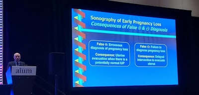
"The key principle for ultrasound in early pregnancy loss is to virtually eliminate false positives," Doubilet said.
 Dr. Peter Doubilet, PhD, talked about ultrasound's use in determining whether a pregnancy is ectopic, as well as the consequences of false-positive and false-negative readings.
Ultrasound is used for patients who present with positive symptoms of pregnancy, with the gestational sac typically being the first object seen about five weeks into a pregnancy. However, abnormal vaginal bleeding is a sign of early pregnancy loss or ectopic pregnancy. Doubilet said that about 25% of early, clinically recognized pregnancies end in miscarriage.
"And those are only the clinically recognized ones, and then there are others that aren't recognized that end in miscarriage," he said.
However, false-positive and false-negative results have consequences, with Doubilet saying that the former is "way more consequential." This is because treatments following false-positive readings for ectopic pregnancies, such as methotrexate, can inadvertently cause miscarriages, stillbirths, or birth defects for healthy embryos.
Doubilet's five rules to consider for fetal ultrasound include the following:
- If a mass is extraovarian, it's "almost certainly" an ectopic pregnancy. If the mass is inside the ovary, it's a corpus luteum.
- If you're unsure about masses in or beside the ovary, press with a transvaginal transducer. If the mass moves with the ovary, it's most likely a corpus luteum. If it moves separately from the ovary, it's an ectopic pregnancy.
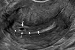
- A gestational sac located in the cervix is likely a cervical ectopic pregnancy, especially if it's well-formed and has a fetal heartbeat. It is likely also a miscarriage in progress if it is a flattened sac without a heartbeat. If you can't tell and the patient is stable, leave the scan for one or two days. The ectopic pregnancy will still be there while the miscarriage in progress will have passed.
- If you have an eccentric gestational sac in the uterus, it's likely to be an interstitial ectopic pregnancy. This also applies if it bulges the uterine contour and has no visible surrounding myometrium. If you're unsure, 3D ultrasound can help provide the correct diagnosis.
- If you see a gestational sac in the uterus on ultrasound, a separate adnexal mass is likely to be a tubal ectopic, heterotopic pregnancy. This also applies if it has a heartbeat or a yolk sac in the adnexal mass in addition to the intrauterine pregnancy. The mass is likely to be a corpus luteum if there is no heartbeat or yolk sac. Heterotopic pregnancies are "very rare" while corpus luteums are common.


