A standardized report template and sonographer training can improve ultrasound’s performance for diagnosing appendicitis in children, according to findings published November 5 in the Journal of Pediatric Surgery.
Researchers led by Alexa Turpin, MD, from Nemours Children’s Health in Wilmington, DE, found that this approach led to less time spent in the emergency department and decreased imaging costs per patient.
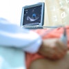
“Abdominal ultrasound should be the first-line imaging for pediatric patients with suspected acute appendicitis,” Turpin and co-authors wrote.
The Pediatric Appendicitis Score (PAS) is a validated tool for diagnosing appendicitis in children. Patients receive a score from 1 to 10 based on the presence or absence of certain signs and symptoms, with scores grouped into categories of low (0 to 3), moderate (4 to 6), and high (7 to 10).
The researchers suggested that the PAS score could be used to create an imaging pathway. While ultrasound is easier to access than MRI and doesn’t use radiation, incomplete visualization can lead to more imaging workup and more time spent in the emergency department.
Turpin and colleagues sought to improve ultrasound’s performance to position the modality for first-line imaging for all patients. In 2022, the team reviewed appendix ultrasound exams performed in 2020. It identified scanning technique and ambiguity in the reporting of findings as major contributors to subpar ultrasound performance. Sonographers underwent training that focused on bowel and appendix imaging.
The team created a standardized template for ultrasound and MRI, adapting the imaging pathway so that all PAS levels underwent ultrasound first. The group focused the study on whether MR imaging was needed after ultrasound and how the pathway affected clinical team time (time from patient being placed in an emergency department room to disposition).
The study included 2,612 children in the pre-intervention phase and 594 in the postintervention phase. The children ranged in age from 7 to 14.
Turpin and colleagues reported the following findings:
The imaging pathway led to a diagnostic ultrasound rate of 56% compared to the baseline rate of 27%.
After adjusting the imaging protocol to an ultrasound-first approach for all patients as opposed to high PAS scores only, the percentage of patients requiring axial imaging after ultrasound did not increase.
Clinical team time decreased from 347.6 minutes to 299.3 minutes, while imaging cost per patient decreased from $6,177.38 to $4,812.30 (p < 0.001 for both).
They noted that despite the decreased clinical team time observed in this approach, the longer times may be due to patients who do not have appendicitis. This is due to these patients likely needing more workup when their imaging results come up negative.
Finally, the investigators added that while the percentage of patients needing multiple imaging modalities on average did not increase due to the pathway, the percentage varied from month to month.
“It will be important to continue to monitor this trend, since patients in the moderate PAS group may be more likely to have equivocal ultrasound and therefore require additional cross-sectional imaging if there remains a suspicion for acute appendicitis,” they wrote.
Read the entire study here.