Interpretation errors by the radiologist occur when a lesion with suspicious features is mischaracterized as a benign or probably benign lesion. This situation may occur because of lack of experience, fatigue, or radiologist inattention.12 Other causes, though, are that the radiologist does not use all the necessary views to assess the lesion characteristics or that the lesion is changing slowly and comparison with older examinations was not made.
When a seemingly circumscribed mass is identified, assessment of its borders should be made with spot compression. A round mass may have margins that are indistinct or microlobulated that become apparent only with spot compression, and these findings warrant biopsy. Microcalcifications require magnification views in order to assess their morphology and number accurately. Decisions about the characteristics of a lesion identified on screening are made based on diagnostic mammographic images and not the screening views alone.
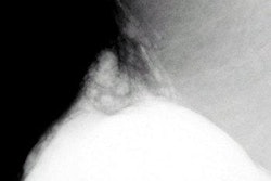
Decisions about lesion location should not be made without verification of the position on multiple views. For example, one should not assume that a lesion overlying the pectoralis muscle in the superior aspect of the MLO view is a lymph node. If its features are not that of a node, additional views are performed to determine its location and significance (figures 8a -- 48KB and 8b -- 57KB). Ultrasound may be a helpful adjunct in the evaluation of a posteriorly located mass seen on one view only.
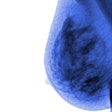
Cancers that present with subtle signs of carcinoma are most challenging to diagnose. Such subtle signs include relatively circumscribed masses, focal asymmetric densities, architectural distortion, and small groups of punctate or amorphous calcifications. Well-circumscribed cancers are very uncommon, but do exist. Swann et al13 found at least a partial halo sign around 25 of 1000 breast cancers. A well-defined mass found on a baseline mammogram is considered to be probably benign and is followed at an early interval; increase in size of a non-cystic circumscribed mass warrants further intervention (figures 9a -- 45KB and 9b -- 46KB).
Asymmetric densities are frequently present on mammography and are associated with a relatively low positive predictive value of malignancy. However, asymmetries that are developing, that are associated with microcalcifications or architectural distortion, or that are palpable are more worrisome and are often biopsied. Invasive lobular carcinoma accounts for approximately 8% to 10% of breast cancers, and is a type of cancer easily missed. Common presentations of invasive lobular carcinoma are an asymmetric density, architectural distortion, or with negative mammography.14
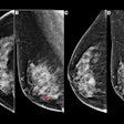
As more women undergo screening mammography on a routine basis, the possibility that a slowly changing cancer goes undetected may increase. The doubling time for breast cancer ranges from 44 to 1,869 days.15 Low-grade malignancies may not change to an obvious degree between annual interval mammograms. Malignant calcifications have been reported to be stable for as long as 63 months. However, comparison with older studies three or more years before may demonstrate the change in size of the lesion. A mammographic abnormality should be judged by its worst features, not its most benign features. Therefore, a lesion that has suspicious features, but seems to be stable for one or even two years, may still need to be biopsied (figures 10a -- 41KB and 10b -- 46KB).
Next: Conclusion & References
1 2 3 4


