Two major causes of missed breast cancers reflect radiologist errors, namely, perception and interpretation problems. In perception errors, the lesion is included in the field of view and is evident, yet the radiologist fails to observe it. The lesion may or may not have subtle features of breast cancer, rendering it less visible than more obvious lesions.
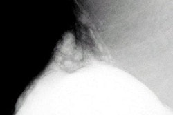
Types of lesions that are more easily not perceived are small non-spiculated masses (figure 1a -- 57KB), (figure 1b -- 57KB), and (figure 1c -- 45KB),areas of asymmetry and architectural distortion, or small clusters of faint amorphous calcifications. Bird et al2 found most cancers were missed because of perception problems, and the common types of lesions missed were noncalcified indistinct or spiculated densities. Goergen et al10 found that missed cancers were statistically significantly of lower density and were seen on only one of two views more often than were detected cancers.
There are several steps that should be taken routinely to avoid missed perception of a lesion. Films should always be placed as mirror images, MLOs together and CCs together. The radiologist's search pattern should include comparing portions of the images side by side to look for any focal density. A suggested search pattern emphasizing a mirror image approach is shown in (figure 2 -- 50KB). This search pattern enhances the ability to identify focal asymmetry density and small masses. The identification of a focal density on one view should prompt a search in the same arc (measured from the nipple) on the other view (figure 3 -- 32KB). The posterior aspect of the breast in the retroglandular region is an area sometimes not observed by the non-expert mammographer, and attention must be applied to this zone specifically (figures 4a -- 42KB and 4b -- 46KB).
Architectural distortion may be the only sign of breast cancer in a dense breast. The parenchyma must be scrutinized for any disruption of the orientation of the elements or for an area of focal pulling or tethering of the tissue (figures 5a -- 36KB and 5b -- 47KB). Architectural distortion must be further evaluated unless it represents a documented post-surgical scar.
The observation of microcalcifications optimally requires careful evaluation of each film with a magnifying glass, as well as the naked eye. Optimal reading conditions necessitate high luminance viewboxes with obscuration of extraneous light. Magnification views should be performed to evaluate questionable faint microcalcifications and the morphology of observed microcalcifications.
Another perception problem that is extremely important in its impact on patient management is the diagnosis of multicentric breast cancer. Multicentric breast cancer (two or more or cancers in more than one quadrant) is a contraindication to breast conservation therapy. The observation of an obvious finding (benign or malignant) may cause the "happy eye syndrome," misleading the radiologist into not looking carefully for other lesions (figures 6a -- 51KB and 6b -- 38KB).
Contralateral synchronous breast cancer occurs in 0.19% to 2.0% of patients,11 so careful attention must also be paid to the opposite breast. When one lesion that is suspicious for cancer is observed, the next step should be the search for other suspicious areas in both breasts that would require biopsy (figures 7a -- 46KB and 7b -- 43KB).
Next: Interpretation errors
1 2 3 4


