Republished with permission from Applied Radiology, October 2000
Although mammography is the gold standard for the detection of early breast cancer, according to data from the Breast Cancer Detection Demonstration Project, the false-negative rate for screening mammography may be as high as 8% to 10%.1 Since then, other authors have suggested an even higher rate (up to 25%) of missed breast cancers on screening mammography.2, 3 Studies have suggested that double reading4-6 or the use of computer-aided diagnosis7, 8 may increase the accuracy of mammographic interpretation. Double reading of screening mammography has been found to improve the sensitivity for breast cancer detection by 5% to 15%.4-6
Causes of missed breast cancers can relate to a variety of factors, including those related to the patient, technical issues, perception, and interpretation. In a review of 320 cases of breast cancer, Bird et al2 found that 24% were missed at screening by one reader. Of the missed cancers, 61% were visible in retrospect and not called, and 18% were interpreted incorrectly. Only 5% were accounted for by technical errors.2 Careful attention to quality control, positioning, comparison with prior studies, and the meticulous use of additional views and adjuvant studies are necessary to minimize the false-negative rate of mammography. This article will discuss the causes of missed cancers and guidelines for reducing the false-negative rate of mammography.
Patient factors
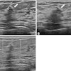
Inherent breast density can certainly compromise the ability to detect a mass, particularly a noncalcified, non-distorting lesion. In the interpretation of the mammogram of a patient with dense parenchyma, particular attention to any areas of architectural distortion and careful assessment for faint microcalcifications are needed. A patient with dense parenchyma, a negative mammogram, and a palpable mass needs ultrasound for further lesion evaluation.
Other patient factors relate to difficulties in positioning and visualizing posterior breast tissue. This may occur in patients who are very tense, but also occurs in patients who have had strokes, who have shoulder problems, and in those who are otherwise debilitated.
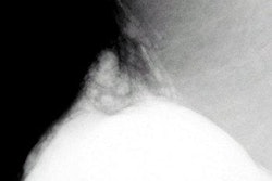
Lesions that are located in areas that are difficult to image with mammography are inherently challenging to diagnose. Lesions located at the chest wall, particularly those high in the upper inner quadrant near the sternum or in the posterior-inferior aspect of the breast, are difficult to visualize.
Technical factors
Proper positioning and image contrast are absolute requirements for mammography. The technologist must adhere to positioning standards in order to maximize the amount of tissue included on the films.9 In order to include an area of palpable abnormality on the images, the examination must be tailored appropriately and the necessary views must be performed. Palpable areas should be identified with radiopaque markers, and the radiologist should verify that any posteriorly located palpable lesion is included in the field of view.
For densities seen on the mediolateral oblique (MLO) view only, a mediolateral (ML) view is helpful to determine if the lesion is real and whether it is located medially or laterally. Lateral lesions project lower on the ML view than on the MLO view, and medial lesions show the reverse displacement. Exaggerated craniocaudal (CC) views are also helpful to demonstrate a posteriorly located lesion seen on the MLO view only. The use of a spot compression view alone may be misleading in the assessment of a density seen on one view only. A true lesion may look less apparent on spot compression as other overlying tissue is displaced from it. Additional off-angle projections (i.e., ML or rolled CC views) should be utilized to complete the evaluation of a possible lesion.
The technologist must optimize image contrast and avoid underpenetrated or overpenetrated images. Proper placement of the photocell is necessary to achieve correct optical density on the images. Careful attention to daily processor quality control is also necessary to optimize contrast.
Next: Perception factors
1 2 3 4

