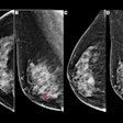
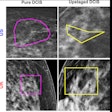
ARLINGTON, VA - Compared to mammography, contrast-enhanced MRI is more accurate for predicting both the diameter and volume of residual tumors in women who received chemotherapy for locally advanced breast cancer, according to research presented at the American College of Radiology Imaging Network (ACRIN) fall meeting.
The results are potentially an important step toward the optimization of surgical outcomes, particularly in women undergoing breast-conserving surgery.
"Mammography diameter did not correlate with residual disease size for lesions overall, or for the subgroups of single mass or single regional enhancements," said Dr. Constance Lehman, Ph.D., professor of radiology at the University of Washington School of Medicine in Seattle.
Using MRI to monitor breast cancer treatment response remains in its infancy, and mammography is also far more commonly used for presurgically assessing residual breast cancer.
Led by Nola Hylton, Ph.D., a professor of radiology at the University of California, San Francisco, along with colleagues at nine centers across the U.S., the ACRIN 6657 protocol is an imaging component within the larger Investigation of Serial Studies to Predict Your Therapeutic Response With Imaging and Molecular Analysis (I-SPY) breast cancer trial network. Lehman unveiled the results on Thursday at the ACRIN fall meeting, and they will also be discussed at the 2009 RSNA meeting in Chicago.
ACRIN 6657 has three primary aims:
- Evaluate MRI's ability to predict disease-free survival after neoadjuvant therapy (follow-up results pending)
- Assess the modality's ability to predict response to such treatment early in therapy (presented at last year's ACRIN and RSNA meetings)
- Evaluate MRI's ability to measure residual disease post-therapy and before surgery, the topic of this week's study
ACRIN 6657 defines breast cancer as an invasive cancer 3 mm or larger without distant metastases. Participants underwent a total of four MRI scans, including one at baseline, a second early in treatment with the chemotherapy drug anthracycline, a third between anthracycline and taxane regimens, and a final MRI scan along with a clinical exam and mammography before surgery to remove residual tumor.
Radiologists measured the longest tumor diameter by MR and mammography, and pathologists measured the longest tumor diameter of residual disease on the tumor specimen, Lehman noted. Signal-enhancement ratios were assessed by computer to measure MRI tumor volume based on enhancement thresholds.
The study estimated residual disease size using Pearson coefficient correlations between the presurgical size measurements and the pathological residual disease measurements.
Of 237 patients enrolled between 2002 and 2006, 215 (97%) were available for this final analysis, representing extraordinary commitment on the part of both participants and investigators "in a very difficult trial to recruit for, and a very intensive trial once they enroll," Lehman said.
The great majority of lesions (71%) presented as masses, followed by nonmasslike disease dubbed "regional enhancements" (23%), and 6% of disease foci that were not characterized.
Pearson correlations of clinical and imaging size for all lesions showed that "the strongest predictor of pathologic longest diameter or residual disease was the final MRI longest diameter measurement," Lehman said. "When we looked at single masses alone that changed, it was the MR volume that was the strongest predictor [r = 0.39, p < 0.0013] of pathologic residual disease."
Presurgical estimation of residual disease for all lesions compared to pathology
|
For single regional enhancements, the final MRI diameter (r = 0.50, p = 0.0028) was the strongest predictor of residual disease.
"We noted that ... the longest diameter at MRI was the best correlate to the pathologic size across all lesions, as well as for the subgroup of single regional enhancements," Lehman said. "However, the MRI volume was the best correlate to pathologic residual disease size for the subgroup of single masses."
For all lesion types, the longest diameter by clinical exam also correlated with the pathologic size (r = 0.37, p < 0.0001), though not as strongly as for the two MRI measurements. This finding warrants further investigation to which the group will respond with various regression analyses, she said. It's also important to note that even though pathologic lesion size is used as the gold standard, it is not 100% accurate, Lehman added.
Compared to MRI, mammography measurements didn't correlate with lesion size at pathology overall, or for the mass or regional enhancements, she said.
"In the majority of programs across the country, mammography is used much more commonly than MRI in assessing regional disease prior to surgery, and we think this finding is important to be aware of," Lehman said.
But even at her own institution, using MRI to follow women through treatment and presurgical assessment is not yet routine.
"We do use the combination of clinical exam and ultrasound, and when there is any question, MR," she said. "I do think that for presurgical assessment of any of the patients planning breast conservation therapy, MRI is the method of choice."
By Eric Barnes
AuntMinnie.com staff writer
October 2, 2009
Related Reading
Patient age, clinical indication need to be considered in breast MRI, September 18, 2009
CAD enhances breast MRI specificity, September 17, 2009
Breast MRI CAD algorithm predicts cancer metastasis, February 6, 2009
Breast MRI pinpoints residual disease after chemotherapy, December 19, 2008
MRI beats mammo for gauging breast cancer treatment response, October 7, 2008
Copyright © 2009 AuntMinnie.com


