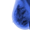
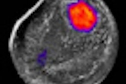
A computer-aided detection (CAD) application that analyzes both full-field digital mammography (FFDM) and dynamic contrast-enhanced MRI images distinguishes between malignant and benign lesions better than traditional single-modality CAD, according to a study published this month in Academic Radiology.
Using contrast-enhanced MRI as a complement to mammography has been shown to improve the accuracy of breast cancer diagnosis, particularly for high-risk women; merging information in CAD programs across these two modalities could have the same effect, according to researchers at the University of Chicago (Acad Radiol, September 2010, Vol. 17:9, pp. 1158-1167).
"Although the results of CAD systems for single imaging modalities [such as mammography, breast sonography, and breast MRI] are encouraging, merging information across different modalities is recently attracting more attention," wrote Yading Yuan, PhD, and colleagues.
Yuan and colleagues constructed a multimodality CAD dataset of 213 lesions (168 malignant, 45 benign) from two larger sets:
- An FFDM database with 432 biopsy-proven lesions (255 malignant, 177 benign), acquired with a Senographe 2000D unit (GE Healthcare, Chalfont St. Giles, U.K.)
- A dynamic contrast-enhanced (DCE) MRI database with 476 lesions (347 malignant, 129 benign), acquired with a 1.5-tesla whole-body Signa MRI system (GE)
Each of the 213 lesions was present on both FFDM and DCE-MRI images, which were acquired between 2002 and 2006.
To analyze the lesions, the team used a CAD scheme developed at the University of Chicago that manually marked lesion location (a particular point on an FFDM image or a region of interest on a DCE-MRI image); the computer then automatically segmented the mass lesions and extracted lesion features. The team used receiver operator characteristics (ROC) analysis to evaluate the performance of certain features in distinguishing between malignant and benign lesions.
"We investigated the performance of a CAD scheme with computerized features extracted from mammography alone, DCE-MRI alone, and the combination of these two modalities," Yuan and colleagues wrote. "Our results demonstrate that combining information from mammography and DCE-MRI yielded significantly higher diagnostic accuracy, in terms of [area under the curve (AUC)] values, than the use of single-modality information only, in the task of differentiating between malignant and benign lesions."
Use of CAD to analyze only mammography features yielded an AUC of 0.74 ± 0.04, while analysis of only DCE-MRI features yielded an AUC of 0.78 ± 0.04. The CAD algorithm performed better when analyzing both FFDM and DCE-MRI together, yielding an AUC of 0.87 ± 0.03, higher than the scores produced by either modality alone.
One of the study's limitations was that the majority of the lesions in the set of 213 were malignant -- an imbalance to be expected, because patients with BI-RADS scores of 3 or greater at mammography are more likely to be referred to MRI, according to the authors. In any case, multimodality CAD could be the next big thing in radiology computer-aided reading.
"Our investigation indicates that multimodality CAD is a promising way to distinguish between malignant and benign lesions," they wrote.
By Kate Madden Yee
AuntMinnie.com staff writer
September 28, 2010
Related Reading
Double readers top CAD in finding breast tissue deformities, July 26, 2010
U.K. study finds mammo CAD can save money, November 9, 2009
Why are correct CAD marks ignored? It's anybody's guess, November 9, 2009
CAD finds breast cancers missed at CR mammography, July 31, 2009
Mammography CAD recalls baffle CADET II researchers, March 20, 2009
Copyright © 2010 AuntMinnie.com


