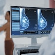
Using a full-field digital mammography system with small detectors to image women with large breasts can result in nearly 50% higher radiation dose compared to imaging women whose breasts are a better match for the detector, according to researchers from Boston.
Dr. Cathy Wells, PhD, led a team of researchers from Beth Israel Deaconess Medical Center who found that women with large breasts imaged on mismatched detectors received higher radiation doses than are recommended by the American College of Radiology (ACR): nearly 5 mGy versus ACR's recommended 3 mGy to 4 mGy for 2D mammography.
"This project originated with the observation that, at our academic center, a subset of mammography patients were receiving more radiation than others -- specifically, women with large breasts who were imaged with a small detector," Wells told attendees at the RSNA 2011 meeting in Chicago. "We wanted to quantify that observation."
Wells pointed out that there are several different types of digital detectors used in various mammography systems, and that these detectors vary in terms of technology and manufacturing protocols. Digital detectors typically come in preset sizes of small and large, which, in turn, "can result in a potential mismatch between a patient's breast size and the mammographic detector size available at the time of imaging," she said.
The study included 864 patients who presented for screening and diagnostic mammography at Beth Israel during a six-week period from November to December 2009. Wells' group gathered data on patient age, breast size (large or small), detector size used, number of views obtained, and the mean glandular dose per breast.
Optimal detector size was determined by viewing prior images or, if those were unavailable, by technologist estimation based on a woman's body size. The researchers calculated mean glandular dose for imaging performed on appropriately matched or mismatched breast-detector pairs.
Screening mammography patients with large breasts imaged on a small detector received a significantly higher radiation dose (4.9 mGy versus 3.3 mGy) and a greater number of views (5.9 versus 4.6), compared with women whose breasts optimally matched the detector size.
Diagnostic mammography patients with large breasts imaged on a small detector also received a higher radiation dose (8.2 mGy versus 6.7 mGy), compared with optimally matched breast-detector pairs. However, these patients did not receive an increased number of views. Compared to patients with large breasts, those with small breasts underwent significantly more views when imaged on a large detector (5.4 versus 3.5).
"A mismatch in breast-detector size results in a significantly greater radiation dose to patients with large breasts imaged on a small detector," Wells concluded. "Patients with small breast sizes did not receive increased radiation when imaged on a large detector, but did receive more views during diagnostic imaging workup, suggesting patient positioning may be more difficult with a mismatch in breast-detector size."
An audience member asked Wells if there are any objective criteria that radiologic technologists can use to determine the correct detector size for a particular woman.
"We collected data on patients' breast size by asking them to report their bra size," Wells responded. "A cup size of C seemed to be the breaking point for those [negatively] affected by radiation dose."


