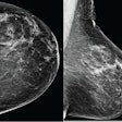
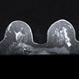
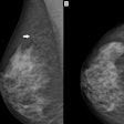
Imaging surveillance is an acceptable alternative to surgical excision in patients with benign papillomas, according to a presentation made at the American Roentgen Ray Society (ARRS) meeting earlier this month.
The papillomas must not have any cell abnormalities when diagnosed at breast core biopsy. This is an uncommon condition, representing only about 1.4% of all imaging-guided core biopsies, according to lead presenter Dr. Jessica Leung of California Pacific Medical Center's Breast Health Center in San Francisco.
The study included 119 papillomas in patients who had image-guided percutaneous core biopsies between January 1996 and October 2011. Of the 119 papillomas, 32 were calcifications and 87 were masses.
For 66 of these lesions, patients were followed for at least two years with imaging and no surgical excision. No cancer was found in this group, according to Leung.
Patients with the remaining 53 lesions underwent surgical excision; 50 of the lesions had benign histology, and three lesions were ductal carcinoma in situ.
The histologic upgrade rate of 2.5% closely approximates the 2% probability of malignancy that is accepted for BI-RADS 3 -- probably benign -- lesions, Leung noted. Because core biopsy reliably excluded cancer in the majority of benign papillomas not associated with atypia or atypical ductal hyperplasia, women could be safely followed using imaging surveillance only, the researchers concluded.


