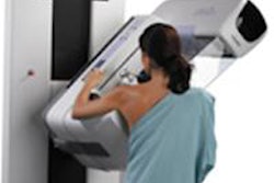
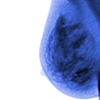
The addition of digital breast tomosynthesis to a screening mammogram increased the rate of breast cancer detection by 11%, according to one of several studies on 3D mammography presented this week at the American Roentgen Ray Society (ARRS) meeting in Washington, DC.
Researchers at Yale University's Smilow Cancer Hospital in New Haven, CT, discovered the added value of tomosynthesis when they retrospectively reviewed a population of 14,684 women who received screening mammograms at Yale between August 2011 through July 2012.
Almost 60% of the women only had a full-field digital mammogram (FFDM, Selenia, Hologic). A total of 42 cancers were detected among these 8,769 patients, for a cancer detection rate of 4.8 per 1,000 women.
A total of 5,915 women received both a mammography and a tomosynthesis exam (Selenia Dimension, Hologic). Thirty-two cancers were identified in this group, for a cancer detection rate of 5.4% per 1,000 women, a rate 11% higher than the 2D rate.
The percentage of invasive and intraductal cancers detected among the two groups was similar, according to scientific session presenter and principal investigator Dr. Jaime Geisel, a breast imager and assistant professor of radiology at Yale University School of Medicine. But tomosynthesis appeared to be more valuable in women with dense breast tissue: 54% of the women whose cancer was found with tomo had dense breast tissue, compared with 21% of those with cancer who only received 2D mammography.
Today, the majority of screening mammograms performed at Yale now include tomosynthesis imaging, Geisel said.
Tomosynthesis and high-risk patients
The study that Geisel presented was one of several made by Yale researchers on breast tomosynthesis at ARRS 2013.
A team led by Dr. Reni Butler, an assistant professor of diagnostic radiology, wanted to assess the potential contribution of tomosynthesis to breast cancer visualization in a high-risk patient population. They reviewed 125 mammographically visible cancers in 107 patients imaged with FFDM and tomosynthesis who had a high risk of having breast cancer.
Seventeen women in this group were classified as high-risk, having either had a very strong family history of breast cancer, were BRCA1 and/or BRCA2 carriers, or had undergone chest radiation as part of treatment for Hodgkin's lymphoma. Cancer visibility was graded by a team of seven breast imagers.
Two of the cancers were only visible on tomosynthesis, and six were more visible on tomosynthesis than on FFDM. All the malignancies that were better seen or only seen on tomosynthesis were either masses, focal asymmetries, or areas of architectural distortion, Butler said. Five of the eight patients in this group had heterogeneously dense breast tissue.
Five of these eight patients were diagnosed with infiltrating ductal carcinoma, and three were diagnosed with infiltrating lobular carcinoma.
"We believe that the addition of tomosynthesis to 2D mammography may improve the visualization of invasive cancers presenting as noncalcified lesions in high-risk women," Butler concluded, and recommend that additional research be undertaken.
2-view tomo recommended
A third study from Yale-New Haven Hospital assessed whether breast cancers could be adequately identified from a single-view screening study, or whether two-view exams were better.
The findings reinforced the current practice in the U.S. of acquiring both craniocaudal (CC) and mediolateral oblique (MLO) views of the breast when performing a combined mammogram and tomosynthesis imaging.
A team of seven breast imagers reviewed tomosynthesis images of 106 patients who had been diagnosed with 115 cancerous breast tumors from their mammography images. Only about half (54%) could be seen equally well on both views, 7% could only be seen on one view but not the other, and 39% were better seen on one view only.
A total of 34% of the cancers were either better or only seen on the craniocaudal view. Lead author and presenter Dr. Noa Beck, a radiologist and breast imaging fellow at Yale School of Medicine, explained that because the CC view achieves better compression, this may have been why this view showed lesions more clearly.
Beck attributed the location in the breast as the primary reason that some cancerous lesions were only seen in mediolateral oblique views.
"We did the study to see if one tomosynthesis view would be sufficient," Beck said. "The study results emphasize that obtaining both views is necessary to ensure that a cancer will be optimally visualized."


