Tuesday, November 28 | 12:15 p.m.-12:45 p.m. | BR234-SD-TUA2 | Lakeside, BR Community, Station 2
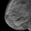
Computer-aided diagnosis of contrast-enhanced spectral mammography (CAD-CESM) can help reduce false positives, according to this Tuesday afternoon poster presentation.A team led by Dr. Bhavika Patel of the Mayo Clinic Hospital in Phoenix sought to evaluate whether CAD-CESM could improve the technology's performance when compared with that of an experienced radiologist. For the study, the group included 50 biopsy-proven lesions found on CESM between August 2014 and December 2015. Histopathological analysis showed 24 benign and 26 malignant cases.
A breast radiologist manually noted lesion boundaries on the different CESM exam views; the team then extracted a set of morphological and textural features from the images to train the CAD tool. Patel and colleagues compared the tool's performance with diagnostic predictions of two other radiologists.
Overall, CAD-CESM accurately identified 45 of the 50 studies, for an accuracy rate of 90%. The detection rate for the malignant group was 88% (three false-negative cases), and the rate for the benign group was 92% (two false-positive cases).
Compared with CAD-CESM, the first radiologist had an overall accuracy of 78% and a detection rate of 92% for the malignant cases (two false negatives) and 62% for the benign cases (10 false positives). The second radiologist had an overall accuracy of 86%, with a detection rate of 100% for the malignant cases and 71% for the benign cases (eight false positives).
The findings show that CAD-CESM could offer radiologists additional decision-making support.
"The results of our feasibility study show potential for a CAD-CESM tool to provide complementary information to radiologists, mainly through potentially reducing the number of false-positive findings on CESM," the group wrote. "Future studies will include more patient cases with image data to train and improve the classification scheme."


