A new mammography technique that uses an artificial intelligence (AI) algorithm to map the biological tissue composition of tumors on dual-energy mammography images could help reduce unnecessary biopsies, according to a study published online December 11 in Radiology.
The findings show promise for the use of radiomics in breast imaging, wrote a team led by Karen Drukker, PhD, of the University of Chicago. Radiomics is an extension of computer-aided diagnosis; it quantifies tumor phenotypes by mining image features from mammography.
 Karen Drukker, PhD, of the University of Chicago.
"In recent years, investigators have shown success using radiomics for breast cancer, extracting a variety of clinically relevant features, merging them into signatures, and estimating the probability of malignancy of identified lesions," Drukker and colleagues wrote.
The researchers investigated whether they could reduce unnecessary biopsies by using radiomic characteristics such as lesion texture, size, shape, and morphology with dual-energy mammography and a new technique called three-compartment breast (3CB) imaging. 3CB imaging was developed by John Shepherd, PhD, from the University of Hawaii in Honolulu. The technique measures water, lipid, and protein tissue composition throughout the breast, which could provide a biological "signature" for the tumor, according to Drukker and colleagues. For example, more water in the tumor tissue suggests angiogenesis, an early sign of cancer development.
"Our study was designed to explore whether the combination of mammography radiomics and analysis of 3CB images derived from dual-energy mammography would improve specificity and reduce unnecessary recalls," Drukker told AuntMinnie.com. "The goal is to have fewer unnecessary biopsies without missing cancers."
For the study, the researchers acquired dual-energy mammograms using the 3CB technique immediately prior to biopsy in 109 women with suspicious breast masses (BI-RADS categories 4 or 5). They evaluated these images using AI algorithms developed by Maryellen Giger, PhD, and colleagues at the University of Chicago.
The combination of 3CB image analysis and mammography radiomics improved the positive predictive value of biopsy performed (PPV3) from 32% for 2D mammography alone to almost 50%, the group found. It also had a 97% sensitivity rate.
| Performance of 3CB imaging and mammography radiomics | |||
| Performance measure | Conventional 2D diagnostic mammography | 3CB mammography | Mammography radiomics plus 3CB |
| Positive predictive value of biopsy performed (PPV3) | 32.1% | 34% | 49% |
| Sensitivity | 100% | 97% | 97% |
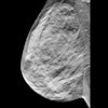
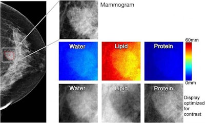 71-year-old woman with 1.6-cm invasive ductal carcinoma (BI-RADS category 5, with category C breast density). Low-energy mammogram and corresponding regions of interest for mammogram (top) and breast tissue composition images (bottom two rows). Courtesy of RSNA.
3CB imaging can be performed with conventional mammography as well as with digital breast tomosynthesis with minimal effect on workflow and only a 10% higher radiation dose, Drukker and colleagues noted.
"The potential exists for wide application of 3CB imaging in diagnostic breast imaging and perhaps also in screening," they wrote.
The technique could also improve patient care, Drukker told AuntMinnie.com.
"This combination of 3CB imaging and mammography radiomics could not only reduce unnecessary biopsies but also patient anxiety and healthcare costs," she said.


